Saturday, 20 May 2017
Vinnie Jones advert for British Heart Foundation - Superb
If some geezer collapses infronta ya, what do you do?
Wednesday, 17 May 2017
Lacerations in the ER Part 1: Before you stitch!
Here are my notes from the 'Advanced wound care' lecture, the last lecture before graduation. After just an hour with this prof I felt like I could handle any laceration that comes into the ER. Hopefully after this post you will feel a similar confidence:
History is important!
Before touching the wound, you should know exactly how it happened, when it happened and what instrument/surface cause the laceration. Its important because wounds heal differently and are devitalised in many different ways depending on these factors.
Location is relevant, the face heals better than the hands. Some areas are more mobile and better perfused.
Patient factors;
Patient factors will affect healing and can be important clues for risk of infection (diabetes), healing time etc.
Steroids slow healing, HIV doesnt affect wound healing, Keloids are common in afro-americans.
Find out the patient's Tetanus Status, you have probably heard about tetanus prone wounds, this is a myth. You can get tetanus from a corneal abrasion. Make you sure you find out about the primary series of vaccinations! no good giving a booster if someone hasn't had their primary series! this is a common situation with immigrants in our ER. Many people forget to ask! CDC advice
The key to good wound practice is IRRIGATION. rememeber: "the solution to pollution is dilution" Water is the best irrigant, you can use plain old tap water, its the volume that matters, taking the patient over to the tap you can supply much more water than a simple syringe from a saline bag. You will never completely eliminate the bacteria, it just needs to have a lower enough concentration so that colonies don't form (sample principle as the lab). Consider the environment, kid falls and hits head on table legvs kid who falls and hits head on road kerb. You need volume and pressure
Sensation:
The gold standard to test sensation is a 2 point discrimination (5mm apart for the hands), make sure there eyes are closed of course. A plastic surgeon can repair most nerves proximal to the DIP joint of the fingers, you will need them to fix any nerve injuries. From a malpractice point of view you need to perform the gold standard.
You can investigate tendon injuries in a variety of ways, a good way to test is to ask the patient to assume the position of function, you will spot any flexor injuries easily.
Position of function: arms raised to shoulder height, with hands pointing up and open chest position. imagine a policeman says "hands up!"
Flexor injuries you should call the surgeon. There is a small no mans land, the deep palmar lacerations. If the palm hurts when they move their hand then they have probably knicked the palmar sheath. With little pain you can probably leave this but a surgical referral is probably best.
Many extensor tendon injuries can be splinted and will heal well.
Remove foreign bodies. 90% of glass can be seen on X-ray
Use anaesthetic, local anaesthesia or nerve block. nerve blocks are very useful and with ultrasound very easy to perform (with practice). Use Lidocaine (short acting) or Bupivicaine (medium acting 8hours half life). Some areas are tender, for example the sole of the foot, do a nerve block!
Allergic reactions to lidocaine itself are impossible! (only to the preservatives used within, which are rare now). You can be almost 100% sure there will be no allergic reaction if you use single use vials or cardiac lidocaine.
There has never ever been a documented case of allergci reaction to cardiac lidocaine.
Most lidocaine reactions are just a vasovagal reaction to the needle ha.
When dealing with the face or kids, topical agents are great eg. Tetracaine. You can wack it on in triage.
Explore all Wounds!
Anaesthesia, Betadine on the neighbouring skin surface (not inside the wound). you are ready to suture!
SUMMARY:
- History of injury
- Location considerations
- Pateint factors
- Tetanus status
- Good neurovascular and functional exam
- Irrigation
- Anaesthesia
 |
| Get ready to stitch this guy up |
Monday, 15 May 2017
An Incredibly Trippy History of the World
I actually learnt so much from this video! Although unbearably trippy it is actually really entertaining and surprisingly accurate. hats off to Bill Wurtz for this creation.
Sunday, 14 May 2017
Quality Improvement in Hospital
These notes are based on a wonderful lecture I had today titled 'quality improvement in health care pathways'. It was a short lecture full of simple but powerful ideas.
I am quite fortunate that my hospital is modern and has a lot of money. One of the ways the administration has decided to spend this money is on a 'quality care' unit whose job is simply to improve patient outcomes and increase the 'quality' of care.
Quality is notoriously difficult in healthcare to measure. The main way that quality is measured in most institutions is by comparison to local and national mortality and infection rates. Yet these gives you no idea about the actual quality of care, it only tells you if more or less people are dying at your hospital compared to another hospital.
Quality is the actual clinical outcome, the treatment of the 'disease' and the patient well being, does the patient leave the hospital in a condition comparable to how he/she was before getting the 'disease'?
A great idea that came out was to use the percentage of patients that returned for follow up as a surrogate for quality, and they are investigating that now.
One way you can indirectly determine quality of care is by using patient forums (groups of patients meeting together), or using patient questionnaires. Many points that are raised by patient forums are simple and often overlooked. One group of patients who had urological operations, noted how they all had no idea what underwear to wear. It may sound stupid to the consultant who is focusing on defeating the cancer being treated (yes the true priority), but the underwear was important to the patient and the source of a great deal of stress.
"If you improve the little things the cancer becomes just a little less important"
The quality care programme at the hospital is a relatively new concept and has taken a very long time to be adopted. Apparently it took about 7 months to get a group of 20 people to talk together, 20 people all involved in different parts of the patients care pathway; nurses, doctors, physiotherapists, radiologists etc.
Communication is a big issue if you want to work on improving quality.
Unfortunately many doctors didn't appreciate advice from the nursing staff at the start, it took a long time for this communication loop to open up. Doctors interpreted the advice as nurses telling the doctors how to do their job, hostile thinking. Just because you’ve done something for 20 years doesn’t mean it is right or couldn’t be better.
Once the problem of communication is solved, drastic changes are seen. The benefits are seen not only among the hospital staff. Patient counselling groups offered to all the patients undergoing prostate cancer resection, were very popular. Patients spoke in a large group with all the staff involved in their care. Hearing about all the people going through the same problem was incredibly enlightening for the patients, even if it regarded embarrassing topics like sexual life after surgery. Understanding the process and what life is like around and after the surgery was helpful. Clinical outcomes were even seen to improve, for example continence after prostate resection was improved. Well prepared patients took their pelvic floor muscle training before surgery seriously, understanding the importance and hearing from other patients.
The quality care team started their approach with observation. They took doctors along and tried to be patients, from the start of the process to the very end. Starting from the parking lot straight away they realised why the little things are important, they couldn't even find the department, there were no signs! We need to take care of patients from the very beginning.
Their are little things you can do that can have a massive impact on patient outcomes and quality of care. Two small examples of changes at our hospital that improved quality of care:
- When a stroke victim is on the way to the hospital emergency department in the ambulance the neurologist is called before and is ready at the door when the patient arrives. as apposed to waiting ten minutes for the neurologist to arrive who has been called only on patient arrival. Time is brain after all. The Dr waits for the patient the patient doesn't have to wait.
- A case manager, who calls patients and organises their follow ups with them on the phone as apposed to the normal "see so and so after six months for ct scan" etc. speaking directly with the patient you can assure they book an appointment and also it fits around them.
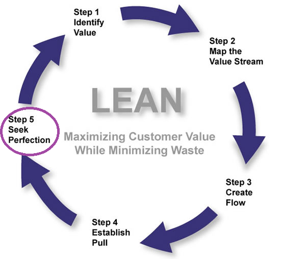
Some of the hospitals ideas came from the famous Toyota LEAN method which you can find out about in this interesting BBC discovery podcast:
http://www.bbc.co.uk/programmes/p042k1bfQuality is the actual clinical outcome, the treatment of the 'disease' and the patient well being, does the patient leave the hospital in a condition comparable to how he/she was before getting the 'disease'?
A great idea that came out was to use the percentage of patients that returned for follow up as a surrogate for quality, and they are investigating that now.
One way you can indirectly determine quality of care is by using patient forums (groups of patients meeting together), or using patient questionnaires. Many points that are raised by patient forums are simple and often overlooked. One group of patients who had urological operations, noted how they all had no idea what underwear to wear. It may sound stupid to the consultant who is focusing on defeating the cancer being treated (yes the true priority), but the underwear was important to the patient and the source of a great deal of stress.
"If you improve the little things the cancer becomes just a little less important"
The quality care programme at the hospital is a relatively new concept and has taken a very long time to be adopted. Apparently it took about 7 months to get a group of 20 people to talk together, 20 people all involved in different parts of the patients care pathway; nurses, doctors, physiotherapists, radiologists etc.
Communication is a big issue if you want to work on improving quality.
Unfortunately many doctors didn't appreciate advice from the nursing staff at the start, it took a long time for this communication loop to open up. Doctors interpreted the advice as nurses telling the doctors how to do their job, hostile thinking. Just because you’ve done something for 20 years doesn’t mean it is right or couldn’t be better.
Once the problem of communication is solved, drastic changes are seen. The benefits are seen not only among the hospital staff. Patient counselling groups offered to all the patients undergoing prostate cancer resection, were very popular. Patients spoke in a large group with all the staff involved in their care. Hearing about all the people going through the same problem was incredibly enlightening for the patients, even if it regarded embarrassing topics like sexual life after surgery. Understanding the process and what life is like around and after the surgery was helpful. Clinical outcomes were even seen to improve, for example continence after prostate resection was improved. Well prepared patients took their pelvic floor muscle training before surgery seriously, understanding the importance and hearing from other patients.
The quality care team started their approach with observation. They took doctors along and tried to be patients, from the start of the process to the very end. Starting from the parking lot straight away they realised why the little things are important, they couldn't even find the department, there were no signs! We need to take care of patients from the very beginning.
Their are little things you can do that can have a massive impact on patient outcomes and quality of care. Two small examples of changes at our hospital that improved quality of care:
- When a stroke victim is on the way to the hospital emergency department in the ambulance the neurologist is called before and is ready at the door when the patient arrives. as apposed to waiting ten minutes for the neurologist to arrive who has been called only on patient arrival. Time is brain after all. The Dr waits for the patient the patient doesn't have to wait.
- A case manager, who calls patients and organises their follow ups with them on the phone as apposed to the normal "see so and so after six months for ct scan" etc. speaking directly with the patient you can assure they book an appointment and also it fits around them.
Some of the hospitals ideas came from the famous Toyota LEAN method which you can find out about in this interesting BBC discovery podcast:
Monday, 8 May 2017
A Brief History of Stroke
Stroke is a leading global cause of death and
disability-adjusted life years (DALYs, see below for definition), second only to ischemic heart disease. The incidence of stroke varies across different countries and
increases exponentially with age.


First defined as ‘apoplexy’ by the
father of medicine Hippocrates himself in 2400BC, it was not until around the
1600’s that the link between the potentially devastating sudden symptoms and
the brain was made (hence the name ‘apoplexy’, which meant in Greek ‘struck
down by sudden violence’). This discovery was made by the documentation of
intracranial haemorrhage in the brains of cadavers who died of stroke by
Johannes Wepfer (Since 2005 the “Johann Jacob Wepfer Award” is given at the
European Stroke Conference for outstanding work in cerebrovascular diseases).


After extensive work in defining the anatomy of the cerebral vasculature by
Thomas Willis in Oxford 1664, and the discovery of an anatomo-pathological
association by the Paris Medical School in the late 1800s, apoplexy became
better known as ‘cerebrovascular accident’. The term ‘stroke’ was a lay term, originating
from the belief the disease was a sort of ‘stroke of gods hand’ or ‘stroke of
justice’, a punishment for wrongdoing or pleasure-seeking. It later became the
definitive name for the disease in 1962 when the chest and heart association
produced a booklet titled ‘Modern Views
on ‘Stroke’ Illness’.
Even at this time, the mid twentieth century, it seemed
doctors still approached stroke with slight nihilism or hopelessness. Up until
1935 bloodletting was the primary therapy for stroke. Vomits, purges and enemas
were all treatments for stroke in the beginning of the nineteenth century, not
so different from Hippocrates own ‘replacement of humours’ before the start of
the millennium. Nothing seemed to improve the prognosis, patients miraculously
recovered, died or faded away with permanent disability; modern medicine had a
long way to go still!
 |
| Bloodletting, 'back in the day' |
In the 1950s doctors began to experiment with angiography,
anticoagulants and surgery for the treatment of stroke. A few years later, team
approaches to stroke patients started in many hospitals with the collaboration
of physiotherapists, nurses, dietitians, surgeons, internists, occupational
therapists, speech therapists and general practitioners. Rehabilitation became
one of the main contemporary treatment responses to stroke. By the end of the
20th century and with birth of stroke associations around the world,
there seemed to be some light in the tunnel, perhaps stroke is curable and
preventable.
Stroke treatment and management has come a long way in the last
fifty years, advancements in angiography and the introduction of aspirin
therapy and intravenous thrombolysis have improved survival massively.
In 2008,
stroke moved from being the 3rd leading cause of death in the USA to
the fourth, it then jumped a further rank to fifth in 2013, a reflection of
accelerating science and improving prognosis!
DALY: The sum of years of
potential life lost due to premature mortality and the years of productive life lost
due to disability. One DALY can be thought of as
one lost year of "healthy" life. The sum of these DALYs across the
population, or the burden of disease, can be thought of as a measurement of the
gap between current health status and an ideal health situation where the
entire population lives to an advanced age, free of disease and disability.
References:
-http://vizhub.healthdata.org/gbd-compare/
Institute for health metrics and evaluation. Accessed 03/04/2017. Images.
-Catherine E. Storey, Hans Pols, Chapter 27 A history of cerebrovascular disease, In: Michael J. Aminoff, François Boller and Dick F. Swaab, Editor(s), Handbook of Clinical Neurology, Elsevier, 2009, Volume 95, Pages 401-415, ISSN 0072-9752, ISBN 9780444520098, http://dx.doi.org/10.1016/S0072-9752(08)02127-1.
-Molnár
Z. Thomas Willis (1621-1675), the founder of clinical neuroscience. Nat Rev
Neurosci. 2004;5(4):329-35.
-Van der worp HB, Van gijn J. Clinical practice. Acute ischemic stroke. N Engl J Med. 2007;357(6):572-9.
-Pound P, Bury M, Ebrahim S. From apoplexy to stroke. Age Ageing. 1997;26(5):331-7.
Friday, 5 May 2017
Monday, 1 May 2017
If BY RUDYARD KIPLING
If you can keep your head when all about you
Are losing theirs and blaming it on you,
If you can trust yourself when all men doubt you,
But make allowance for their doubting too;
If you can wait and not be tired by waiting,
Or being lied about, don’t deal in lies,
Or being hated, don’t give way to hating,
And yet don’t look too good, nor talk too wise:
If you can dream—and not make dreams your master;
If you can think—and not make thoughts your aim;
If you can meet with Triumph and Disaster
And treat those two impostors just the same;
If you can bear to hear the truth you’ve spoken
Twisted by knaves to make a trap for fools,
Or watch the things you gave your life to, broken,
And stoop and build ’em up with worn-out tools:
If you can make one heap of all your winnings
And risk it on one turn of pitch-and-toss,
And lose, and start again at your beginnings
And never breathe a word about your loss;
If you can force your heart and nerve and sinew
To serve your turn long after they are gone,
And so hold on when there is nothing in you
Except the Will which says to them: ‘Hold on!’
If you can talk with crowds and keep your virtue,
Or walk with Kings—nor lose the common touch,
If neither foes nor loving friends can hurt you,
If all men count with you, but none too much;
If you can fill the unforgiving minute
With sixty seconds’ worth of distance run,
Yours is the Earth and everything that’s in it,
And—which is more—you’ll be a Man, my son!

Sunday, 30 April 2017
Dr John Hinds Bike Crash Prehospital Care and Emergent Thoractomy
I have been looking for the best medical documentaries, and while searching the r/documentaries setion on reddit I found a great post linking the video below. While not entirely a documentary per se it is a super interesting lecture on the specifics of bike crash prehospital care. He is a fantastic lecturer with a great sense of humour, well worth a worth.
Dr Hind died while responding to an accident at the 2015, Skerries Road Races, in County Dublin, Ireland, RIP.
Another lecture by Dr Hind, this time on Emergent Thoractomy and more cases from the races.
Dr Hind died while responding to an accident at the 2015, Skerries Road Races, in County Dublin, Ireland, RIP.
Another lecture by Dr Hind, this time on Emergent Thoractomy and more cases from the races.
Thursday, 27 April 2017
Twitter Conference: Charing Cross Vascular Symposium #cx2017 Day Two
Since my paper is being presented and I am unable to be there, I will be 'attending' the Charing Cross Vascular Symposium through the wonderful medium of twitter. Following the hashtag #CX2017 I hope to catch as much as possible here on my blog. This is what I caught from Day 2:


 thanks to the twitter warriors: @cookEVAR @claudicant @perealtes @ozvascdoc @veryanmed1 @torbjornlundh @cxsymposium @vascularMD

Again the organisers continued with there fantastic ask the audience sections

And the results were interesting...


Wednesday, 26 April 2017
Twitter Conference: Charing Cross Vascular Symposium #cx2017 Day One
Since my paper is being presented and I am unable to be there, I will be 'attending' the Charing Cross Vascular Symposium through the wonderful medium of twitter. Following the hashtag #CX2017 I hope to catch as much as possible here on my blog.


— BIBA MedTech (@BIBAMedTech) April 25, 2017

The first day was all about peripheral artery disease and the audience were asked some interesting questions regarding the their views on PAD, here are the results:

Dr Hauton alongside Dr Mastracci with her wonderful video in my last post addressed concerns on radition exposure, pointing out that 1 DSA image is roughly equal to 500 fluoro images:

Mr Weinberg posted the following, pointing out that PAD patients receive better outcomes when combining exercise with revascularisation (linkhere):

Lucky man Dittmar Bockler loves his DynaCT:

What about smoking cessation in vascular patients, try try again:

Dr Anders Wanhainen on the need for a disease specific solution for type B dissection.

Currently watching closely the twitter-web today, following todays focus on Abdominal aortic aneurysms... watch this space.
Thanks to @PereAltes @Angiologist @CXsymposium @CookEVAR @ozvascdoc
Tuesday, 25 April 2017
Dose Awareness Matters
New video produced by the Vascular surgeons at the Royal Free Hospital in London. Right now it is the Charing Cross vascular symposium one of the biggest vascular surgery conferences in the world. Stay connected by following the hashtag #cx2017
Sunday, 23 April 2017
Saturday, 22 April 2017
Friday, 21 April 2017
The Obesity Paradox in Type 2 Diabetes
Summary of a guest lecture given today by Dr P Costanzo an Interventional radiologist working in the UK. You can find him on pubmed here and on twitter here:
Increasing BMI has been shown to increase all cause mortality in this NEJM study in 1.46 million white patients. Further extrapolation showed this was mainly due to cardiovascular death.
Furthermore obesity levels are on the rise. So too are the levels of type 2 diabetes. However, the mortality levels for patients with type 2 diabetes has not shown the same rise infact it has been petty stable for many years.
A large cohort of type 2 diabetic patients (T2DM) was taken by Costanzo et al. and divided into categories of weight and then following up over 10 years for mortality. The kaplan meier survival curve was interesting, displaying the so called obesity paradox. Patients who were underweight with a low BMI (less than 20 or 18.5) had the highest mortality. Increasing BMI showed a protective effect with the highest BMI values having the lowest mortality. A paradox indeed.
Further extrapolation of the data by cause of death showed a protective effect of obesity in T2DM in sepsis and cancer (again paradoxical, considering cancers relationship with obesity).
Dr Costanzo went on to explain possible mechanisms of this, citing the important relationship between low birth weight and increased lifetime risk of T2DM. And how this may be part of the so called evolutionary Thifty phenotype, a phenotype in which high blood sugar can be maintained in starvation providing a survival advantage. It is well known that subsaharan populations (and also indian populations) who move to say the UK (or anywhere) and start a western diet are likely to develop diabetes. He mentions the lipgenic model of T2DM and how subcutaneous fat is neutral to us but visceral fat is the fat that as it accumulates increases cardiovascular mortality. There is a kind of tipping point where when lets say SC fat is full, visceral fat begins to accumulate (where is the level?).
The last part of Dr Costanzos talk was incredibly interesting. HB1AC levels documented across all values of BMI is more or less the same in his cohort (unreleased data unfortunately, paper release in 2017), except for the underweight BMI values in which it generally higher. You can postulate that HB1AC is therefore not linked to mortality, and infact other studies confirm this. My notes run out at this point, I guess I was trying to concentrate, but the final part of this section he shows that good BP control in T2DM can reduce mortality in T2DM and perhaps glycemic control has little to do with cardiovascular mortality.
In fact a nice point was that two new anti-diabetic drugs being studied at the moment; Liraglutide and Empagliflozin, are the only drugs that have been shown to reduce cardiovasuclar mortality in T2DM. These two drugs also have a blood pressure lowering effect.

Monday, 10 April 2017
Saturday, 8 April 2017
My 2017-2019 Foundation Programme (UK) Application Experience
Almost a year after starting the process, I finally got my foundation programme (FP) placement on Thursday. I am dead excited to start work as a junior doctor, seriously cannot wait! I thought I would do a quick breakdown here of how the process went, so maybe some future applicants can have a better idea.
Some Background:
-International undergraduate from Italy
-Fourth Decile class rank (This doesnt tell you anything about the level of clinical skill just how awesome your class is, :P)
-Previous degree
-A single publication (nothing groundbreaking, but many a late night spent writing and researching)
Eligibility (August 2016):
In order to start the whole process the FP office had to make sure I was eligible. This eligibility process required identity documentation and a deans statement from my university (basically a statement saying I have enough clinical experience from my degree for the programme). I also had to sit the IELTS english exam in June in order to be eligible for the programme. This was all done in the summer 2016 (before foundation programme allocation applications began).
My IELTS academic score average was 8.
October 2016:
In October the applications began properly and I submitted evidence of my degree and all my personal data. I also had to find two references, a clinical one (my mentor) and academic one (dean of the school who knew me well).
My final EPM score was 44 (not bad)
Within that application in October I had to rank the UoAs (the different regions of England). I ranked the UoAs in the following order:

December 2016: I then sat the SJT (situational judgement test) on the first sitting in December. I flew in from Italy and stayed in Brighton the night before. Unfortunately, I ate the dodgiest salad at the LEON restaurant in Brighton centre (sorry to call them out, but there was no doubt that that salad was dodgy). After explosive vomiting all night, and no sleeping a wink, I got the train to London the next morning for the exam, dehydrated and pale as milk. Sat the exam and staggered home.
My final SJT score 36.66 (awful score considering the studying I did, brilliant score considering the physical state I was in ha).
Finished the entire exam without leaving any questions, reviewed about half my answers before time was up.
Beginning of March 2017: Got into South Thames Foundation school!! Fantastic!
Due to the fact that South Thames foundation school is so large (over 800 placements in total), they decided to split the area into six STHAM groups. I had to do an additional ranking of the six STHAM groups. Due to the lack of crossover between the two cities I had the hard choice of choosing between London or Brighton. I really wanted to go to Kings College for the liver transplant unit and London for other reasons so I ranked the groups like this:

End of March 2017: Got into London Links 2 area, woohoo!
Then I had to rank my programme preferences. There were 152 in total to rank. In the end I ranked about 110 of them. I realised at this point I had probably made a big mistake, having got my second choice of STHAM group I would probably have a low decile compared to the other applicants for entering the programme I wanted. (In fact I checked the STFS website and the decile averages for each STHAM were released, I was in the last deciles, sad face).
Ranking all the programmes is difficult and you have to decide what you want to priotize, the location or the rotations (London or surgery, which is more important?). You also want a good variety in your programme without any lets say 'wasted' or repeated rotations.
I spoke to a bunch of friends who had gone through the programme and this was their advice:
- Don't do emergency medicine in the first year, do it in the second year
- Avoid highly specialised rotations for example like opthalmology (sorry eye guys, no offense)
- GP rotations are nicer in the summer
- Get as much ward time as possible
- Rotations are more important than location but try and get at least one year in a big teaching hospital
Spending about two days on excel with the list of placements, I ordered everything based on the advice above and with two main priorities:
Had to contain Surgery
Had to have Emergency Medicine in F2
After finding all these rotations, I then ranked them in order of location, prioritising placements at Kings College or Guys hospitals (which I'm sure everyone did).
(There were about 40/50 of these, the rest I literally ranked based on the awesomeness of rotations).
The excel file I created kind of looked like this after colour coding and moving around everything a million times.
 |
| (looks like a mess right, but I had a system) |

Last Thursday (April 2017): I found out my placement! F1 Maidstone F2 Croyden! Cannot wait!
Pretty damn chuffed, the placementwas my 17th ranked placement.
So from today there is still lots to do, mainly issues now regarding provisional licensing and transfer from Italy to England. I'll keep y'all posted.
 |
| (Pretty sweet ey, look forward to many blog posts relating to these topics) |
(Disclosures: All the information is regarding my own application and I do not believe I am revealing any information without consent).
Friday, 7 April 2017
Pancreas Transplantation BMJ State-of-the-Art Review Summary
(Another medical student orientated summary of a recent review, this time Pancreas Transplantation.)
Successful pancreas transplantation can result in durable glycemic control and improved survival for patients with diabetes. There seems to be no other treatment in medicine that has the same improving success rates over time and is being applied less and less (the number of pancreas transplants performed in the US has decreased every year during the past decade). In other words, more patients could probably benefit from pancreas transplantation than currently undergo the procedure.
Most people are diagnosed with type 2 diabetes, with type 1 diabetes accounting for 8-10% of all diabetes cases.
In the UK 3.5 million people are diagnosed with diabetes, with approximately 0.5 million still to be diagnosed. The incidence is increasing.
First successful pancreas transplant was in 1966, at the University of Minnesota.
The number of transplants increased steadily until 1996.
Survival at this point (1996) was 91% at one year and 84% at three years.
The introduction of ciclosporin in the 1980s dramatically increased survival, further efficacy of transplant was enhanced with introduction of tacrolimus and mycophenolate in the 1990s.
Between 2005 and 2014 pancreas transplantation number decreased by 20%. Reasons for this decline were probably; improved medical management of diabetes, decline in organ donor quality (more obese and old), lack of consistent referral of transplant candidates from endocrinologists.
Three main pancreas transplantation types:

Success rates of pancreas transplantation have improved with time likely due to increasing experience with these complex patients.
UK current survival rates SPK five year survival 88%, Pancreas only transplants five year survival 78%.
No real studies have directly compared the costs of pancreas transplant vs conventional medical therapy but there have been theoretical models that concluded that SPK is the most cost effective strategy after accounting for varying probabilities of patient and graft survival.
Because pancreas transplantation can also establish normoglycemia it is reasonable to infer that this intervention would also improve or stabilise end organ complications (eg. retinopathy, nephropathy).
Complications:
Diabetic nephropathy (a microvascalur complication of diabetes) is one of the most important complications of diabetes.
Single centre studies with small cohorts have suggested that pancreas transplantation has a beneficial effect on secondary complications of diabetes.
Data is limited on the long term complications, have been reports of increased infections and hematologic cancers after transplantation.
Quality of life:
QoL improved rapidly after transplantation (measured at four months), the effect did however flatten out later. A minority had decreased QoL emphasising the importance of pre-transplant education to establish realistic expectations for the patient.
Clinical trials:
No multicentre trial has been designed to truly evaluate the true efficacy of transplant compared to best medical therapy in type 1 diabetes.
Islet transplantation (ITA):
ITA is less invasive.
Has good short term results but five year insulin Independence rate are around 11%, despite this these patients achieved avoidance of hypoglycemia and near normal glucose control.
Comparison of ITA vs PTA; PTA has higher morbidity, authors of mentioned study concluded that ITA produces similar outcomes to PTA.
Artificial pancreas:
A closed loop system with a subcutaneous sensor that transmits glucose measurements to an external insulin pump that deliver insulin subcutaneously when needed.
Addition of glucagon in the future could prevent hypoglycemia.
The use of such devices requires the patient reaches a certain level of understanding.
Results from international diabetes closed-loop trial conducted on real patients will be out in 2019.
Future directions:
Need the development of biomarkers to diagnose rejection and monitor patient immune status.

This summary was for the following paper: http://www.bmj.com/content/357/bmj.j1321
(all the information and images were from the above paper).

Successful pancreas transplantation can result in durable glycemic control and improved survival for patients with diabetes. There seems to be no other treatment in medicine that has the same improving success rates over time and is being applied less and less (the number of pancreas transplants performed in the US has decreased every year during the past decade). In other words, more patients could probably benefit from pancreas transplantation than currently undergo the procedure.
Most people are diagnosed with type 2 diabetes, with type 1 diabetes accounting for 8-10% of all diabetes cases.
In the UK 3.5 million people are diagnosed with diabetes, with approximately 0.5 million still to be diagnosed. The incidence is increasing.
First successful pancreas transplant was in 1966, at the University of Minnesota.
The number of transplants increased steadily until 1996.
Survival at this point (1996) was 91% at one year and 84% at three years.
The introduction of ciclosporin in the 1980s dramatically increased survival, further efficacy of transplant was enhanced with introduction of tacrolimus and mycophenolate in the 1990s.
Between 2005 and 2014 pancreas transplantation number decreased by 20%. Reasons for this decline were probably; improved medical management of diabetes, decline in organ donor quality (more obese and old), lack of consistent referral of transplant candidates from endocrinologists.
Three main pancreas transplantation types:
- PAK = pancreas after kidney transplant (the main role of this type is avoid the morbidity and mortality asociated with dialysis therapy; patients with type 1 diabetes have at least a 33% mortality in teh first five years after starting dialysis).
- PTA = pancreas transplantation alone (has higher rates of technical graft loss and acute cellular rejection, however a very small number of this type are performed, no no reports have rigorously studied the efficacy or quality of life benefits)
- SPK = simultaneous pancreas kidney transplant (most common type of pancreas transplant, typically both organs from the same donor)
Success rates of pancreas transplantation have improved with time likely due to increasing experience with these complex patients.
UK current survival rates SPK five year survival 88%, Pancreas only transplants five year survival 78%.
No real studies have directly compared the costs of pancreas transplant vs conventional medical therapy but there have been theoretical models that concluded that SPK is the most cost effective strategy after accounting for varying probabilities of patient and graft survival.
- To date there have been no randomised controlled trials comparing the different forms of pancreas transplantation against for example intensive insulin therapy, islet transplantation.
- However many single centre studies and registry analyses suggest that pancreas transplantation provides a net benefit compared to kidney transplant alone for patients with both diabetes and chronic kidney disease.
- More controversial is the impact of pancreas transplantation on patient survival in patients with diabetes and preserved renal function. One analysis of transplant registry data reported a survival disadvantage for PAK and PTA recipients.
Because pancreas transplantation can also establish normoglycemia it is reasonable to infer that this intervention would also improve or stabilise end organ complications (eg. retinopathy, nephropathy).
Complications:
Diabetic nephropathy (a microvascalur complication of diabetes) is one of the most important complications of diabetes.
Single centre studies with small cohorts have suggested that pancreas transplantation has a beneficial effect on secondary complications of diabetes.
Data is limited on the long term complications, have been reports of increased infections and hematologic cancers after transplantation.
Quality of life:
QoL improved rapidly after transplantation (measured at four months), the effect did however flatten out later. A minority had decreased QoL emphasising the importance of pre-transplant education to establish realistic expectations for the patient.
Clinical trials:
No multicentre trial has been designed to truly evaluate the true efficacy of transplant compared to best medical therapy in type 1 diabetes.
Islet transplantation (ITA):
ITA is less invasive.
Has good short term results but five year insulin Independence rate are around 11%, despite this these patients achieved avoidance of hypoglycemia and near normal glucose control.
Comparison of ITA vs PTA; PTA has higher morbidity, authors of mentioned study concluded that ITA produces similar outcomes to PTA.
Artificial pancreas:
A closed loop system with a subcutaneous sensor that transmits glucose measurements to an external insulin pump that deliver insulin subcutaneously when needed.
Addition of glucagon in the future could prevent hypoglycemia.
The use of such devices requires the patient reaches a certain level of understanding.
Results from international diabetes closed-loop trial conducted on real patients will be out in 2019.
Future directions:
"Pancreas transplantation stands at a crossroads—without a systematic approach to the procedure and its outcomes, transplant volumes, especially those for PTA and PAK, may continue to decline and the procedure take second stage to therapies such as islet transplantation and closed loop insulin and glucagon delivery systems..... a more systematic approach to characterizing the successes and limitations of pancreas transplantation is needed."Need to develop a uniform definition of graft failure. The most common definition of graft failure at the moment is the requirement of exogenous insulin therapy.
Need the development of biomarkers to diagnose rejection and monitor patient immune status.
UK guidelines:
In the UK, patients with the following conditions are considered
for pancreas transplantation135:
• Pancreas transplantation alone or islet
transplantation alone: patients with severe
hypoglycemic unawareness but normal or near
normal renal function
• Simultaneous pancreas and kidney transplantation
or simultaneous islet and kidney transplantation:
patients with renal failure and insulin dependent
diabetes
• Pancreas after kidney transplantation or islet after
kidney transplantation: patients with functioning
kidney transplants and diabetes.
Most patients who are considered have type 1 diabetes
but some patients with insulin dependent type 2 diabetes
may also be suitable candidates.
This summary was for the following paper: http://www.bmj.com/content/357/bmj.j1321
(all the information and images were from the above paper).

Wednesday, 5 April 2017
NEJM Maternal Immunization Review 2017 Summary
(A medical student orientated summary of a recent NEJM review)
Most childhood vaccines do not start providing adequate protection until the infant is several months old. The immunity gap between birth and this time can be addressed by maternal immunization.
Sex hormones modify immune responses:
Current recommendations are for pregnant women to have influenza and Tdap (tetanus-diphtheria-acellular pertussis) maternal immunisations
Influenza vaccine:

Most childhood vaccines do not start providing adequate protection until the infant is several months old. The immunity gap between birth and this time can be addressed by maternal immunization.
Sex hormones modify immune responses:
- Increase in estradiol is associated with increased Th2 responses and reduced Th1 immune responses.
- Increased progesterone levels is associated with reduced immune response in general
- Overall phagocytic responses, alpha-defensin expression, neutrophil, monocyte and dendritic cell numbers may be increased in 2nd and 3rd trimester in general during preganacy.
- (may explain suboptimal responses to viral infections such as influenza in pregnancy)
- pregnancy is not a generalised state of immunosuppression.
Current recommendations are for pregnant women to have influenza and Tdap (tetanus-diphtheria-acellular pertussis) maternal immunisations
Influenza vaccine:
- Few low income countries regularly vaccinate pregnant women against influenza
- A substantial burden of illness among pregnant women is attributable to seasonal influenza
- The efficacy within infants after birth ranged from 30% to 63% (good for mom, ok for baby)
- There may be potential for protection against adverse birth outcomes, two studies detected a difference in low birth weight however the others did not. One study was sufficicnetly powered to detect a difference but the promising result was offset perhaps by the overall low baseline birth weight. So perhaps the vaccine is more useful as a protection against adverse outcomes.
- influenza infection is associated with an increased rate of subsequent bacterial infection particularly pneumococcal disease. (in fact a substantial proportion of deaths during the 1918 flu epidemic were probably due to strep.pneumoniae).
- use of maternal vaccine and infant vaccine together showed better results than infant vaccine alone in prevention of respiratory illnesses with fever and medically attended acute respiratory illnesses.
Pertussis vaccine:
- Young infants have a disproportionately high burden of severe pertussis in the population.
- studies on the pertussis vaccine have shown high effectiveness and a reassuring safety profile (there were no increaes in adverse birth or pregnancy outcomes).
- There was a slightly higher rate of chorioamnionitis but the authors explained this as perhaps due to practice of labelling fevers as chorioamnionitis to protect from litigation in the USA where the study was conducted
- There is concern though that the vaccine may reduce the immungenicity of the infant DTP (diphtheria-tetanus-pertussis) vaccine. With studies suggesting this, however the clinical relevance is uncertain.
Maternal vaccines in development include vaccines against RSV (respiratory syncytial virus) and GBS (group B streptococcus).
RSV vaccine:
- RSV is the leading cause of viral acute lower respiratory tract illness and the highest morbidity is among preterm infants. Most deaths due to RSV occur in infants
- 2-3% of all neonatal deaths are attributable to RSV.
- Vaccine is needed due to the high burden of RSV infection, particularly among young infants.
- Several RSV vaccines are in development, targeting the RSV F and G proteins mainly.
- Given that preterm infants are a high risk group, recommendation for the gestational age of vaccination will have to take into account adequate antibody transfer for preterm infants.
- In the 1960s a formalin inactivated form of the vaccine against RSV for children was researched. It led to an 'enhanced RSV disease', and increased rates and severity of RSV lower respiratory illness. Thought now to be due to a lack of protective antibodies being produced with an increase in CD4+ priming in the absence of CD8+ cells. This abherrent vaccine slowed further research into RSV vaccines.
- The benefit of a maternal vaccination is that it would bypass immunologic events that would lead to an enhanced RSV disease in infants.
Group B strep vaccine:
- Early onset GBS infection occurs in neonates younger than 7 days and is characterised by sepsis without a focus, pneumonia or meningitis.
- Late onset GBS infection occurs in infants who are 7 to 89 days of age and is characterised with higher rates of meningitis.
- Invasive GBS infection in pregnant women is associated with stillbirth
- GBS is transmitted from colonised mothers during birth (hence why there is universal maternal screening with intrapartum antibiotic prophylaxis).
- One in five pregnant women have evidence of GBS rectal or vaginal colonisation at 23-26 weeks.
- Screening with prophylaxis programmes mentioned above have resulted in reductions in early-onset GBS disease but NO reduction in late onset disease.
- A maternal GBS vaccine could help reduce the burden of GBS disease, particularly late-onset disease in infants.
- there is a trivalent vaccine in development that covers serotypes Ia, Ib and III. The global disease burden includes serotypes II and V, therefore a vaccine will need to be developed still to cover against also these subtypes
Incorporating maternal vaccines into antenatal care has been a challenge in many locations, with the maternal influenza vaccination rate in the US estimated to be around 50%.
Link to paper: http://www.nejm.org/doi/full/10.1056/NEJMra1509044
(all the points above and images were taken from the paper linked above)

Sunday, 2 April 2017
Michael Debakey: The Janitor and I, Saving Lives Together

Source: http://www.freibergs.com/resources/articles/leadership/10-things-we-can-learn-from-the-worlds-greatest-surgeon/ (its a great post and worth a read, it certainly makes a young doctor feel inspired).In one particular encounter, DeBakey began chatting with an elderly janitor who was sweeping the floor. DeBakey asked the man about his wife and children. He told the older man, obviously not for the first time, that the hospital couldn’t function without the janitor because germs would spread, increasing the chances of infection in the hospital. Later in the day, our colleague tracked down the janitor and asked him, “What exactly do you do? Tell me about your job.” With pride, the janitor replied: “Dr. DeBakey and I? We save lives together.”He’s right. After all, consider what would happen to our healthcare systems if the cleaning crews went on strike. DeBakey understood that showing the janitor exactly how he contributes to a larger, more heroic cause is crucial. This creates a powerful dynamic. Realizing that he is working toward a worthy goal, the janitor’s perceptions about his work changed. It had new meaning and his enthusiasm for the job was rejuvenated.
Sunday, 26 March 2017
Friday, 24 March 2017
Summary: WHO Recommendations for Surgical Site Infection Prevention (December 2016)
Surgical site infections (SSIs) are one of the most preventable of all healthcare associated infections,. Prevention is complex and relies on many factors, an integration of a wide range of measures before during and after surgery. In fact SSIs are the most frequent healthcare associated infection in low income countries (second in Europe and USA). Below are the current recommendations from the World Health Organisation that are all aimed at reduced SSIs.

- Don't discontinue immunosuppressive medications before surgery. It can induce a flare of disease activity and interruption of therapy may induce anti-drug antibodies. There is low quality evidence that there may be a reduction in SSIs with anti-TNF drug discontinuation only.
- Give multiple nutrient-enhanced nutritional formulas to underweight patients undergoing major surgery.
- Patients should bathe or shower before surgery with either plain or antimicrobial soap. Either type of soap can be used based on the evidence.
- Patients with known nasal carriage of Staphylococcus Aureus should recieve intranasal applications of mupirocin 2% ointment. The ointment should be applied nasally twice daily for five days. S.aureus carriage is a risk factor for SSIs. There is evidence for effectiveness of this for only cardiothoracic and orthopedic procedures but the panels suggests for all types of surgery. The ointment is only for KNOWN carriers to prevent unnecessary treatment and resistance spread.
- Adult patients undergoing colorectal procedures should have preoperative antibiotics combined with mechanical bowel preparation (MBP) prior to surgery. The use of MBP reduces the intraluminal faecal mass and therefore bacterial load, alongside further bacterial load reduction with antibiotics. The potential harms of MBP should be considered; patient discomfort, electrolyte abnormalities, dehydration, acute phosphate nephropathy. Most common antibiotic regime in the evidence was aminoglycosides combined with anaerobic coverage (metronidazole).
- Hair removal should be with clippers NOT with shaving. This is because microscopic trauma to the skin can increase SSI risk.
- Surgical antibiotic prophylaxis should be done within 120 minutes before skin incision. The evidence showed increased SSIs if the antibiotics were given after skin incision or before 120minutes (note the evidence was of low quality though). Antibiotics with a short half life such as cefazolin or cefoxitin should be given closer to the skin incision time e.g. less than 60min before.
- Surgical hand preparation is vitally important! scrubbing should be done with either antiseptic soap or an alcohol based hand rub (ABHR). There was no difference between povidone-iodine or chlorohexidine. ABHR reduced the number of colony forming units more than antiseptic soap.
- Alcohol based antiseptic solution based on chlorhexidine-gluconate should be used for skin preparation. Alcohol solutions were shown to be more effective than aqueous solutions. Although alcohol based solutions should not be used on neonates and caution should be exercised to prevent contact with eyes, mucosa or accumulation for its flammable nature. Chlorhexidine should absolutely be kept away from the brain, meninges, eyes and middle ear.
- Antimicrobial sealants should not be used. There was no benefit for their use in the literature in preventing SSIs.
- Adult patients undergoing general anaesthesia with endotracheal intubation should receive an 80% fraction of inspired oxygen intraoperatively and if feasible in the immediate postoperative period for 2-6 hours. Infected tissue tends to have a lower oxygen tension than tissue that is non-infected, perhaps due to enhanced oxidative killing by neutrophils. In the postoperative period this benefit was observed with use of a high-flux mask and when normovolemia and normothermia was maintained.
- Normothermia should be maintained with the use of warming devices during the procedure. There are many adverse effects from a hypothermic state (<36degrees); increased cardiac complications, impaired coagulation, impaired wound healing, decreased drug metabolism, decreased immune metabolism and increased SSI incidence.
- Intensive perioperative glucose control. Using an intensive protocol reduced the SSIs although neither an optimal target glucose concentration or timing of control coudl be defined by thepanel. Blood glucose must be closely monitored and hypoglycemia avoided at all costs.
- Sterile disposable or reusable drapes and gowns should be used (Duh!). Although interesting the panel says plastic adhesive incise drapes should be avoided something we use regularly in my unit hmm (apparently they don't effect SSI incidence at all, yeah maybe they dont effect skin edge infection rates but im sure they can help prevent graft infection rates if you are using non native prothesis or grafts, a question for another time, let me find out).
- Wound protector device use should be considered. These devices, small clips that cover the wound edge have been associated with significantly reduced risk of SSI.
- You should irrigate the incisional wound with an aquaous povidone-iodine solution before closure. It removes cellular debris and dliutes possible contamination, results were heterogenous, favouring wound irrigation slightly.
- High risk wounds should be treated with prophylactic negative-pressure wound therapy (pNPWT). PNPWT was shown to significantly reduced SSIs compared to conventional dressings in abdominal and cardiac high risk insicions. A high risk incision is one with poor tissue perfusion, decreased blood flow, hematoma or obvious dead space.
- Antibiotic coated/triclosan coated sutures should be used. Evidence showed that ticlosan coated sutures had a significant effect in reducing SSIs, although there was a statment over possible study limitations due to industry sponsorship.
- Laminar airflow ventilation has no benefit compared to conventional operating room ventilation and shouldn't be used to reduce SSIs.
- Antibiotic prophylaxis should not be prolonged for presence of a wound drain and drains should be removed when clinically indicated. drains can adversly effect the outcomes by affecting anastomotic healing and propagating infections.
- Advanced dressings should not be used over standard surgical dressings for purpose of SSI prevention. There was no evidence that advanced dressings (e.g. silver impregnated) had any benefit over standard gauze dressings.
- Surgical antibiotic prophylaxis (SAP) should not be prolonged after the operation. Most guidelines recommend a maximum SAP duration of 24 hours, infact a single preoperative dose might be non inferior. Surgeons are regularly documented prolonging SAP, this is unnesscary and risks increasing antibiotic reisstance rates. Author does note that prolonging SAP may have a benefit in cardiac and orthognathic surgery only.
The links to the papers where all this was taken from:
Friday, 17 March 2017
Burns
Over 175000 patients visit the emergency department every year with a burns injury in the UK. Burns can be life threatening in the acute phase and severely affect quality of life in the chronic phase with scarring and sometimes even loss of limbs and body parts. Care in the first hours can have a massive impact on the long term outcome, so here is the facts on burns!
FIRST AID:
If you approach a burns patient outside of hospital adopt the SAFE approach first always.
SAFE: Shout for help, Assess scene (is it safe?), ensure its Free from danger before you approach, Evaluate the casualty (ABCDE)). Pay particular attention to the A for Airway in your primary and secondary surveys and look for signs of inhalation injury (listed below).
SAFE: Shout for help, Assess scene (is it safe?), ensure its Free from danger before you approach, Evaluate the casualty (ABCDE)). Pay particular attention to the A for Airway in your primary and secondary surveys and look for signs of inhalation injury (listed below).
After you have done a general assessment of the patient with ABCD, remove any clothing and jewellery around the injury unless they are stuck to the wound, then leave it.
Manage the burn with the 3 C's:
COOL CALL COVER
Cool the burn with normal running tap water for at least 20 minutes, the water should be around 15 degrees. Cooling is beneficial up to three hours after the burn. The rule is to cool the burn but warm the patient! so make sure the rest of the patient is well covered up, you must prevent hypothermia at all costs. Don't use ice.
Call an ambulance.
Cover the injury, use loose clingfilm, on the face you should use wet gauze instead.
EXAMINATION AND HISTORY:
The severity of a burn is judged by the percentage total body surface affected (%TBSA) and the depth of the burn.
3 methods for Judging %TBSA:
- Wallaces rule of nines (adults, picture below)
- Lund and Bowder chart (accounts for age differences)
- Number of hands (Area of palmar surface of hand is equal to1%)
Include all burnt areas in the surface area but NOT very superficial burns, with no blistering and only red and dry.

Judging depth of burn, four levels of depth:
(-Superficial (dry, red and painful, normal capillary refill)
-Superficial dermal (erythematous, small blistering, moist, painful, brisk capillary refill)
-Mid-dermal (dark pink, blistered, sluggish capillary refill, dull sensation)
-Deep-dermal (blotchy red, may be blisters still, no refill, no sensation)
-Full thickness (white or black, eschar often present, no refill and insensate)
This system has largely superceded the degrees system but is roughly the same:
superficial dermal=first degree burn
mid-dermal and deep dermal= second degree burn
full thickness= third degree burn
note that deep dermal and full thickness burns will not heal!
You should pay pay aprticular attention to signs of impending airway obstruction; hoarseness, stridor, snoring and smoke induced inhalation injury which can result in airway oedema and obstruction. So look for signs of burns to mouth, nose, face, singed nasal hairrs, carbonacous sputum.
If any of the burns are circumferential in nature, they could act as a tourniquet with scarring and tissue swelling underneath. This could lead to limb ischemia or even respiratory compromise if the chest wall is involved. You need to look out for this! To preserve the limb the patient may need an 'echarotomy' a surgical incision of the burnt tissue to allow the tissue to expand.
If any of the burns are circumferential in nature, they could act as a tourniquet with scarring and tissue swelling underneath. This could lead to limb ischemia or even respiratory compromise if the chest wall is involved. You need to look out for this! To preserve the limb the patient may need an 'echarotomy' a surgical incision of the burnt tissue to allow the tissue to expand.
FLUIDS:
Loss of water from the burnt area and generalised oedema caused by systemic inflammation can cause a life threatening hypovolemia and organ failure.
Start fluids with all burns greater than 15% TBSA using m-Parkland formula below
and aim for a urine output of 0.5ml per kg body weight.
Remember inhalation burns also lead to fluid losses! (you cannot see the extent of internal burns).
The Modified Parkland Formula:
-give 3-4ml Hartmanns solution per kg body weight per %TBSA over 24hours (half given over first 8hours, over half given over next 16hours).
Dilutional hyponatremia is common and so is hyperkalemia with extensive muscle damage. Electrocution burns can cause rhadbomyolysis and myoglobinuria. You may need to increase the fluid resuscitation to prevent acute tubular necrosis from kidney myogobin overload.
MANAGEMENT and pearls:
Inhalation injury management:
-establish patent airway early and consider intubation early. oxygenate and ventilate. Get arterial blood gases and CO levels.
Get tetanus status
Gastroparesis is common and you should consider inserting a NGT.
Patients are often in pain and emotional distress so give IV analgesia early. Consider opioids as first line (titrated to effect).
Avoid antiseptics and dress wounds with non-adherent dressings and gauze, applie covering bandge very loosely. In a first aid setting clingfilm works very well.
-Paraffin gauze and silver sulfadiazine cream, covered with gauze and bandage.
Consider non-accidental injury and abuse, (does the pattern of injury fit the story).
Antibiotics not indicated in early care
Children: start fluids with 10% TBSA burns and aim for urine output of 1ml per kg body wt.
When to refer to a burns specialist/burns centre:
- Any chemical or electrical burn
- Any burn to face, perineum/genitalia, hands and feet
- TBSA greater than 25% second degree burn
- All full thickness burns
Main complications of full thickness/third degree burns:

- Infections, sepsis
- Tetanus
- Hypothermia
- Hypovolemia
Chemical burn management:
-Brush off any powder first, flush the area with copious amounts of water for 20-30minutes. Note duration of exposure and whether chemical is acid or base.
Surgical management:
(generally by a plastic surgeon or burns specialist)
Dressing, Escharotomy, Escharectomy, Flaps, Graft
-late escharectomy has better aesthetic outcomes but early escharotomy may be necessary for circulation or ventilation problems.

Sources:
-Emergency medicine secrets 6th edition Dr Vincent Markovchick 2016
-Student BMJ January 2016 volume 24
-Lecture notes Prof Klinger Humanitas University 2016 and seminar 2015
Subscribe to:
Posts (Atom)