Monday 31 October 2016

Pericardiocentesis, you stick a needle where?!

Some brief notes of pericardial effusion

Presentation:
The most common presentation is Dyspnea and Tachypnea
In the later stages the patient may even end up arresting with PEA (pulseless electrical activity)

Signs:
Classical becks triad:
Jugular venous distension
Distant heart sounds
Hypotension
Other signs include: Pulsus paradoxus (drop of SBP of greater than 10mmhg on inspiration), low voltages on ECG, electrical alterans, cardiomegaly on CXR.

Risk factors:
Metastatic cancer, mediastinal radiation, end stage renal disease, recent surgery, tuberculosis
This list is very similar to the list of etiologies:
Malignancy, radiation, uremia, dialysis, infection, idiopathic, iatrogenic, post AMI.

Diagnosis:
Clinical diagnosis, best with ultrasound.
On ultrasound you may see; a dilated IVC without changein size on respiration, right ventricle collapse (in fact you may see collapse of any of the chambers).

Treatment:
Pericardiocentesis, depending on ultrasound findings and the expertise of operator, most often performed in the subxiphoid position, with a spinal needle aimed at 45 degrees towards to the left shoulder. Ultrasound is used to guide a spinal needle (keep the needle lateral to the probe so it is always in view, the same plane) into the pericardial sac, avoiding the myocardium. A guidewire and catheter can be positioned to facilitate the drainage. In an emergency setting the procedure can be performed blind or with the help of ECG lead attached to the needle (if you see ST segment elevation then you have gone too far). 
Complications of pericardiocentesis: pneumothorax, coronary artery injury, liver or stomach injury, dysrhythmias, 

Sunday 30 October 2016

Acute Renal Failure and Acute Kidney Injury (ARF vs AKI)

If you look up the definition of acute renal failure (ARF) you will probably find this; "an abrupt and sustained decrease in renal function". Thats all very well but what do any of these words mean?! how abrupt? how long is sustained? renal function measured how? Whats more, over 35 different definitions of ARF are used in the literature with varying mortality and incidence rates. This post should hopefully clear everything up about ARF and AKI and give some tips about how to manage ARF and AKI.

First of all, scrap the term ARF, the term AKI is used now and reflects much better the fact that small decrements in organ function not resulting in organ failure are still clinically important! ARF is used for the last stage of AKI where the kidney actually fails and RRT (renal replacement therapy, for example hemodialysis) is needed. 

Diagnosis:
How do you know if a patient has AKI? look at serum creatinine and urine output!
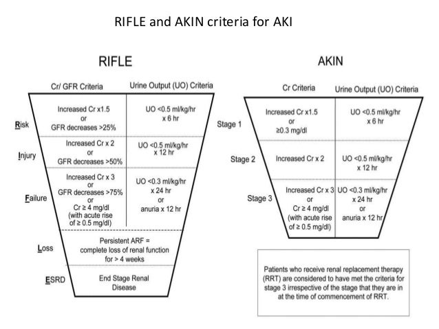
There are two main ways for classifying AKI, the RIFLE criteria and the AKIN criteria, both perform equally well in studies, but I will use AKIN criteria because it is used by the KDIGO guidelines which most of this post is based on.


Picture above shows the AKIN criteria in the red box, as you can see you can divide AKI into three stages depending on the level of serum creatinine (sCr) or urine output (UO) (or directly place them into stage 3 if they are on RRT or anuric for greater than 12 hours). Stage 3 AKI is synonymous with ARF. 

So you have assessed your patients sCr and UO and find that he/she has an AKI. what do you do next?

AKI Management:
  • monitor diuresis (bladder catheterise if not already catheterised)
  • careful physical exam (pay attention to whether the patient is 'wet or dry', you should be careful giving fluids in a 'wet' pt, see later notes on pulmonary edema)
  • monitor fluid balance
  • arterial blood gases
  • order labs: sCr, Na+, K+, Ca2+, Cl-, CBC, urine dipstick, BUN, 
  • renal US (you are looking for the easy dx of obstruction, see etiology below)
  • CXR (pulmonary edema?)
  • avoid contrast agents if at all possible
  • consider ICU if stage 2 and up
  • avoid hyperglycemia
  • careful drug review
  1. discontinue nephrotoxic drugs (eg. vancomycin, gentamycin)
  2. discontinue drugs that impair GFR autoregulation (NSAIDs, ACEi, ARBS(angiotension receptor blockers))
  3. adjust dosage of drugs undergoing renal excretion (many antibiotics)
  4. withhold exogenous potassium (look for K+ containing infusions (such as isolyte) that the patient may be on) and stop potassium sparing diuretics like spironolactone and eplerenone. 
AKI treatment:
Treatment is mainly directed at the underlying causes, for example if the cause is dehydration give fluids, if the cause is haemorrhage consider giving blood transfusion etc. immunosuppressants for vasculitis, discontinue drugs, relieve urinary tract obstructions....
note: No specific treatment for AKI actually exists but we have to manage all the complications well

AKI causes:
The causes of AKI are best divided into three main catergories
  • pre-renal
  • intrinsic (parenchymal)
  • post-renal
Pre-renal AKI:
Basically anything that causes a drop in blood pressure low enough that the kidneys own auto-regulation is unable to preserve renal function. consider:
Volume loss: hemorrhage, dehydration, diarhhea, polyuria, burns
Sequestration of fluids (3rd spacing): pancreatitis, peritonitis, rhabdomyolysis
Blood pressure drop: any form of Shock, hypotensive medications 
(note that not all patients have the same capacity to autoregulate their renal filtration)

Intrinsic AKI:
diseases that affect the kidney directly, consider:
Arteries: thrombosis, embolic events
Pre-glomerular arterioles: Vasculitis, malignant hypertension, atheroembolism, DIC, eclampsia
Glomeruli: glomerulonephritis, thrombotic microangiopathy
Tubulo-interstitium: Acute tubular necrosis (ATN), crystalluria, cast nephropathy, contrast agents
(note that anything that can cause a sustained hypotension can damage the renal epithelium and cause ATN)

Post-renal AKI:
This is caused by an obstruction to the urinary tract, consider:
Bladder outlet obstruction: BPH, urethra stenosis, neurologic bladder
Ureter obstruction in pts with 1 kidney: stones, cancer, papillary necrosis

AKI Complications:
AKI patients can die of four main causes:
  • Hyperkalemia
  • Metabolic acidosis
  • Fluid accumulation (pulmonary edema)
  • Uremic syndrome
Hyperkalemia occurs when serum K+ is >5 mmol/l
Symptoms of hyperkalemia include: intestinal colic, diarhee, weakness/paralysis, arthymias
Hyperkalemia has a distinct ECG: flattened P waves, wide QRS, peaked T waves
(peaked T waves is the first feature to appear)

Metabolic acidosis is caused by the failure of tubular interstutium to excrete normal daily acid load. symptoms include: nausea and vomiting, abdominal pain, hyperventilation, hypotension
(note that acidosis may worsen hyperkalemia)

Fluid accumulation is quite often iatrogenic! careful with hydration. 

Uremic syndrome has many presentations and may cause: pericardial effusion, nausea and vomiting, malaise, confusion, seizures, non specific diffuse abdominal pain, ileus, a tendency to bleed (so called 'lazy' platelets). 

Sunday 16 October 2016

Mean Arterial Pressure (MAP)

I'm currently studying shock, its a huge subject and incredibly important topic in medicine. It doesn't matter what kind of doctor you are, you need to be able to deal with shock. MAP is very relevant in the state of shock.

MAP a great indicator of the perfusion pressure of the organs. Its kind of like an average blood pressure, so if its low there isn't much blood reaching the vital organs. As we all know, blood pressure has two components a systolic and diastolic component. So its not so easy to work out MAP or 'average' blood pressure.

There are some formulas that help (DBP = diastolic blood pressure, SBP = systolic blood pressure):

MAP = DBP + 1/3 (DBP-SBP)

MAP = 2/3 DBP + 1/3 SBP

There are many and quick search of wikipedia or on google will easily come up with many different formules, the two above are the simplest. (note: DBP - SBP is also called the Pulse Pressure).

A normal MAP in a healthy subject is around 90mmHg or in the range 70-110mmHg,
In the treatment of shock we are trying to get the MAP to around 65mmHg or maintaining it there.
(this is because a MAP above 60mmHg is considered enough to perfuse the organs. thus below 60mmHg you should start worrying about organ ischemia).

Monday 10 October 2016

Tissue Factor and Thromboplastin

Tissue Factor (TF) (aka Factor 3 or CD142) is a protein with a key role in coagulation. TF is present in large concentrations in subendothelial tissue of vessels (so by smooth muscle cells and fibroblasts). The endothelium itself can also express TF but only in imflammatory states, same for circulating monocytes.
TF is responsible for triggering the so called extrinsic pathway of coagulation (or TF pathway as its also known). This path is triggered when the vessel is damaged and there is exposure of the underlying TF. It acts as a receptor for Factor 7 which leads to the cleavage of Factor 10 and the activation of the common pathway of coagulation and thrombin activation.

Thromboplastin is the name of a lab reagent and is actually the combination of TF and phospholipids. TF and phospholipids together can trigger coagulation. Partial thromboplastin is just the phospholipids by themselves and can trigger the intrinsic pathway of coagulation (TF not needed to activate intrinsic pathway). So when an 'activated partial thromboplastin time' APTT test is order it is a measure of the intrinsic path of coagulation.

Picture highlighting TF (yellow box), the extrinsic pathway (red box) and common pathway of coagulation (green box). #medicine #coagulation #labs #tissuefactor #bleeding #medicalschool #medicalblog #medED #FOAMed #study

Wednesday 5 October 2016

Fever Of Unknown Origin (FUO)

I am sure you have all watched at least one episode of House, the series in which a witty Hugh Laurie is presented with difficult to solve cases almost every day. Obviously in reality these kind of cases are incredibly rare, but there is one kind of 'difficult to solve' problem in medicine which appears far more often than you think, the febrile illness without an obvious origin, FUO.

Definition and Diagnosis:
Fever greater than 38.3 degrees on several occasions, persisting without diagnosis for at least 3 weeks despite of at least 1 week investigation in hospital. (Later updated to 3days Inpatient investigation or 3days of Outpatient investigation).

Can be further classified into:
Classical (as defined above)
Nosocomial (the fever was absent on admission to the hospital)
Neutropenic (Patient is neutropenic as well, less than 500 neutrophils per mm3)
HIV associated (Patient has confirmed HIV infection)

You cannot conclude a patient has a FUO until you have performed the following basic investigations:
History, Physical exam, Complete blood count, Blood Cultures, Complete metabolic panel and Liver function tests, Urinanalysis and culture, Chest X-ray.

Etiology:
Three catergories of illness can cause FUO; 
infections, malignancies and connective tissue disorders.
There is a long list of the possible causes (See image) but don’t forget Drug Fevers in which a fever can be the sole feature of an adverse drug reaction (most commonly with antibiotics).

Age is very important when considering the etiology, for example in younger patients infections will be much more common (in children around one third of FUO are caused by self limited viral illnesses) and in the elderly (haematological malignancies and solid tumours will be much more common).
Also neutropenia associated FUO will be much more likely linked to a bacterial infection (although never forget genetic neutropenia exists such as cyclic neutropenia and benign familial neutropenia).
Malaria and respiratory infections are a common cause in returned travellers.

First steps:
Re do history and physical exam, a careful history is critical for diagnosis! 
Ask about: animal exposure, immunosupression, drugs and toxins, localising symptoms (for example, jaw claudication is consistent with giant cell arteritis, nocturia with prostatitis etc.). 
Note that the degree of fever, nature of fever curve and response to antipyretics has no specificity to guide the diagnosis. 

After a careful history the following exams will be useful (obviously performed in a targeted nature, guided by your suspiscions):

LEVEL 1 testing:
ESR, CRP, LDH, TST/IGRA, HIVab/RNA, 3 blood cultures for separate sites, Rheumatoid factor, creatine phosphokinase, heterophile antibody test, antinucleaur antibodies, serum protein electrophoresis, procalcitonin can be helpful
LEVEL 2 testing:
CT abdomen, CT chest (if these turn out to be negative move to FDG-PET, although be aware of its high false positive rate)
Level 3 testing: 
Biopsies and Endoscopies

Treatment:
Treatment should be withheld as long as possible until the cause of the fever is determined and empirical antibiotic treatment is not appropriate! However you must obviously consider the patients condition and febrile neutropenic patients have a much higher percentage of bacterial infections and so empiric treatment can be appropriate after cultures have been obtained.

Summary:
  • Most cases of FUO are due to unusal represantations of common diseases rather than exotic diseases
  • Reassess the patient frequently and the don’t underestimate the importance of a very careful history and physical exam (it is in fact critical).
  • Almost any infective agent can be responsible for FUO
  • Look out for Malignancy red flags in the history and physical exam such as symptoms (night sweats, weight loss, pruritus, rectal bleeding), radiation exposure, cigarette smoking, lymphadenopathy, hepatosplenomagaly, petechiae.


A long list of the possible causes of FUO #fever #medicine #fuo #medED #study #medicalschool