Showing posts with label MedED. Show all posts

Friday, 24 March 2017

Summary: WHO Recommendations for Surgical Site Infection Prevention (December 2016)

Surgical site infections (SSIs) are one of the most preventable of all healthcare associated infections,. Prevention is complex and relies on many factors, an integration of a wide range of measures before during and after surgery. In fact SSIs are the most frequent healthcare associated infection in low income countries (second in Europe and USA). Below are the current recommendations from the World Health Organisation that are all aimed at reduced SSIs.

  1. Don't discontinue immunosuppressive medications before surgery. It can induce a flare of disease activity and interruption of therapy may induce anti-drug antibodies. There is low quality evidence that there may be a reduction in SSIs with anti-TNF drug discontinuation only. 
  2. Give multiple nutrient-enhanced nutritional formulas to underweight patients undergoing major surgery.
  3. Patients should bathe or shower before surgery with either plain or antimicrobial soap. Either type of soap can be used based on the evidence. 
  4. Patients with known nasal carriage of Staphylococcus Aureus should recieve intranasal applications of mupirocin 2% ointment. The ointment should be applied nasally twice daily for five days. S.aureus carriage is a risk factor for SSIs. There is evidence for effectiveness of this for only cardiothoracic and orthopedic procedures but the panels suggests for all types of surgery. The ointment is only for KNOWN carriers to prevent unnecessary treatment and resistance spread. 
  5. Adult patients undergoing colorectal procedures should have preoperative antibiotics combined with mechanical bowel preparation (MBP) prior to surgery. The use of MBP reduces the intraluminal faecal mass and therefore bacterial load, alongside further bacterial load reduction with antibiotics. The potential harms of MBP should be considered; patient discomfort, electrolyte abnormalities, dehydration, acute phosphate nephropathy. Most common antibiotic regime in the evidence was aminoglycosides combined with anaerobic coverage (metronidazole). 
  6. Hair removal should be with clippers NOT with shaving. This is because microscopic trauma to the skin can increase SSI risk. 
  7. Surgical antibiotic prophylaxis should be done within 120 minutes before skin incision. The evidence showed increased SSIs if the antibiotics were given after skin incision or before 120minutes (note the evidence was of low quality though). Antibiotics with a short half life such as cefazolin or cefoxitin should be given closer to the skin incision time e.g. less than 60min before. 
  8. Surgical hand preparation is vitally important! scrubbing should be done with either antiseptic soap or an alcohol based hand rub (ABHR). There was no difference between povidone-iodine or chlorohexidine. ABHR reduced the number of colony forming units more than antiseptic soap. 
  9. Alcohol based antiseptic solution based on chlorhexidine-gluconate should be used for skin preparation. Alcohol solutions were shown to be more effective than aqueous solutions. Although alcohol based solutions should not be used on neonates and caution should be exercised to prevent contact with eyes, mucosa or accumulation for its flammable nature. Chlorhexidine should absolutely be kept away from the brain, meninges, eyes and middle ear.
  10. Antimicrobial sealants should not be used. There was no benefit for their use in the literature in preventing SSIs. 
  11. Adult patients undergoing general anaesthesia with endotracheal intubation should receive an 80% fraction of inspired oxygen intraoperatively and if feasible in the immediate postoperative period for 2-6 hours. Infected tissue tends to have a lower oxygen tension than tissue that is non-infected, perhaps due to enhanced oxidative killing by neutrophils. In the postoperative period this benefit was observed with use of a high-flux mask and when normovolemia and normothermia was maintained. 
  12. Normothermia should be maintained with the use of warming devices during the procedure. There are many adverse effects from a hypothermic state (<36degrees); increased cardiac complications, impaired coagulation, impaired wound healing, decreased drug metabolism, decreased immune metabolism and increased SSI incidence. 
  13. Intensive perioperative glucose control. Using an intensive protocol reduced the SSIs although neither an optimal target glucose concentration or timing of control coudl be defined by thepanel. Blood glucose must be closely monitored and hypoglycemia avoided at all costs. 
  14. Sterile disposable or reusable drapes and gowns should be used (Duh!). Although interesting the panel says plastic adhesive incise drapes should be avoided something we use regularly in my unit hmm (apparently they don't effect SSI incidence at all, yeah maybe they dont effect skin edge infection rates but im sure they can help prevent graft infection rates if you are using non native prothesis or grafts, a question for another time, let me find out). 
  15. Wound protector device use should be considered. These devices, small clips that cover the wound edge have been associated with significantly reduced risk of SSI. 
  16. You should irrigate the incisional wound with an aquaous povidone-iodine solution before closure. It removes cellular debris and dliutes possible contamination, results were heterogenous, favouring wound irrigation slightly.
  17. High risk wounds should be treated with prophylactic negative-pressure wound therapy (pNPWT). PNPWT was shown to significantly reduced SSIs compared to conventional dressings in abdominal and cardiac high risk insicions. A high risk incision is one with  poor tissue perfusion, decreased blood flow, hematoma or obvious dead space. 
  18. Antibiotic coated/triclosan coated sutures should be used. Evidence showed that ticlosan coated sutures had a significant effect in reducing SSIs, although there was a statment over possible study limitations due to industry sponsorship. 
  19. Laminar airflow ventilation has no benefit compared to conventional operating room ventilation and shouldn't be used to reduce SSIs.
  20. Antibiotic prophylaxis should not be prolonged for presence of a wound drain and drains should be removed when clinically indicated. drains can adversly effect the outcomes by affecting anastomotic healing and propagating infections.  
  21. Advanced dressings should not be used over standard surgical dressings for purpose of SSI prevention. There was no evidence that advanced dressings (e.g. silver impregnated) had any benefit over standard gauze dressings. 
  22. Surgical antibiotic prophylaxis (SAP) should not be prolonged after the operation. Most guidelines recommend a maximum SAP duration of 24 hours, infact a single preoperative dose might be non inferior. Surgeons are regularly documented prolonging SAP, this is unnesscary and risks increasing antibiotic reisstance rates. Author does note that prolonging SAP may have a benefit in cardiac and orthognathic surgery only. 
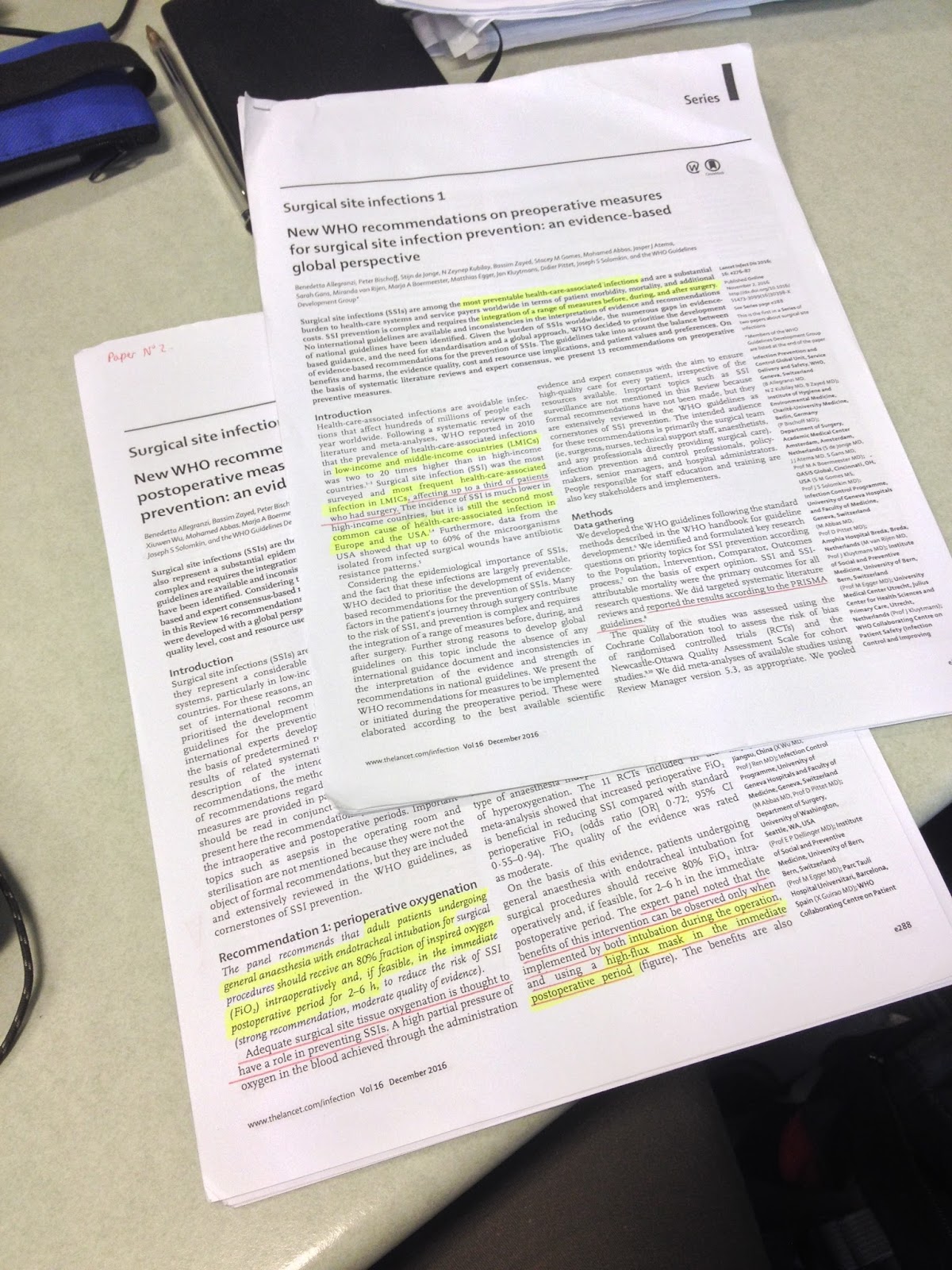
The links to the papers where all this was taken from:

Friday, 17 March 2017

Burns

Over 175000 patients visit the emergency department every year with a burns injury in the UK. Burns can be life threatening in the acute phase and severely affect quality of life in the chronic phase with scarring and sometimes even loss of limbs and body parts. Care in the first hours can have a massive impact on the long term outcome, so here is the facts on burns! 

FIRST AID:

If you approach a burns patient outside of hospital adopt the SAFE approach first always.
SAFE: Shout for help, Assess scene (is it safe?), ensure its Free from danger before you approach, Evaluate the casualty (ABCDE)). Pay particular attention to the A for Airway in your primary and secondary surveys and look for signs of inhalation injury (listed below).

After you have done a general assessment of the patient with ABCD, remove any clothing and jewellery around the injury unless they are stuck to the wound, then leave it. 
Manage the burn with the 3 C's:
COOL CALL COVER
Cool the burn with normal running tap water for at least 20 minutes, the water should be around 15 degrees. Cooling is beneficial up to three hours after the burn. The rule is to cool the burn but warm the patient! so make sure the rest of the patient is well covered up, you must prevent hypothermia at all costs. Don't use ice. 
Call an ambulance. 
Cover the injury, use loose clingfilm, on the face you should use wet gauze instead. 

EXAMINATION AND HISTORY:

The severity of a burn is judged by the percentage total body surface affected (%TBSA) and the depth of the burn.

3 methods for Judging %TBSA: 
- Wallaces rule of nines (adults, picture below)
- Lund and Bowder chart (accounts for age differences)
- Number of hands (Area of palmar surface of hand is equal to1%)
Include all burnt areas in the surface area but NOT very superficial burns, with no blistering and only red and dry.


Judging depth of burn, four levels of depth:
(-Superficial (dry, red and painful, normal capillary refill)
-Superficial dermal (erythematous, small blistering, moist, painful, brisk capillary refill)
-Mid-dermal (dark pink, blistered, sluggish capillary refill, dull sensation)
-Deep-dermal (blotchy red, may be blisters still, no refill, no sensation)
-Full thickness (white or black, eschar often present, no refill and insensate)

This system has largely superceded the degrees system but is roughly the same:
superficial dermal=first degree burn
mid-dermal and deep dermal= second degree burn
full thickness= third degree burn
note that deep dermal and full thickness burns will not heal!

You should pay pay aprticular attention to signs of impending airway obstruction; hoarseness, stridor, snoring and smoke induced inhalation injury which can result in airway oedema and obstruction. So look for signs of burns to mouth, nose, face, singed nasal hairrs, carbonacous sputum. 

If any of the burns are circumferential in nature, they could act as a tourniquet with scarring and tissue swelling underneath. This could lead to limb ischemia or even respiratory compromise if the chest wall is involved. You need to look out for this! To preserve the limb the patient may need an 'echarotomy' a surgical incision of the burnt tissue to allow the tissue to expand. 

FLUIDS:

Loss of water from the burnt area and generalised oedema caused by systemic inflammation can cause a life threatening hypovolemia and organ failure.
Start fluids with all burns greater than 15% TBSA using m-Parkland formula below
and aim for a urine output of 0.5ml per kg body weight.
Remember inhalation burns also lead to fluid losses! (you cannot see the extent of internal burns).

The Modified Parkland Formula:
-give 3-4ml Hartmanns solution per kg body weight per %TBSA over 24hours (half given over first 8hours, over half given over next 16hours).

Dilutional hyponatremia is common and so is hyperkalemia with extensive muscle damage. Electrocution burns can cause rhadbomyolysis and myoglobinuria. You may need to increase the fluid resuscitation to prevent acute tubular necrosis from kidney myogobin overload.

MANAGEMENT and pearls:

Inhalation injury management:
-establish patent airway early and consider intubation early. oxygenate and ventilate. Get arterial blood gases and CO levels.

Get tetanus status

Gastroparesis is common and you should consider inserting a NGT.

Patients are often in pain and emotional distress so give IV analgesia early. Consider opioids as first line (titrated to effect).

Avoid antiseptics and dress wounds with non-adherent dressings and gauze, applie covering bandge very loosely. In a first aid setting clingfilm works very well.
-Paraffin gauze and silver sulfadiazine cream, covered with gauze and bandage.

Consider non-accidental injury and abuse, (does the pattern of injury fit the story).

Antibiotics not indicated in early care

Children: start fluids with 10% TBSA burns and aim for urine output of 1ml per kg body wt.

When to refer to a burns specialist/burns centre:

  • Any chemical or electrical burn
  • Any burn to face, perineum/genitalia, hands and feet
  • TBSA greater than 25% second degree burn
  • All full thickness burns
Main complications of full thickness/third degree burns:

  • Infections, sepsis
  • Tetanus
  • Hypothermia
  • Hypovolemia
Chemical burn management:
-Brush off any powder first, flush the area with copious amounts of water for 20-30minutes. Note duration of exposure and whether chemical is acid or base. 

Surgical management:
(generally by  a plastic surgeon or burns specialist)
Dressing, Escharotomy, Escharectomy, Flaps, Graft
-late escharectomy has better aesthetic outcomes but early escharotomy may be necessary for circulation or ventilation problems. 


Sources:
-Emergency medicine secrets 6th edition Dr Vincent Markovchick 2016
-Student BMJ January 2016 volume 24
-Lecture notes Prof Klinger Humanitas University 2016 and seminar 2015

Friday, 18 November 2016

How do I study for this?

Studying in my final year of medicine, there is one question I get asked by the years below me all the time. "how do you study for this? what should I study from, the books, pubmed or the internet?"
Compared to twenty-thirty years ago and with the boom of the internet we have a huge amount of resources at our disposal. And its this huge variety in educational resources makes this question an increasingly common one. Here is my answer:

Disclaimer: First of all, everyone has their own best method, some people learn best from videos, others from quizzes and case studies, this is an important disclaimer when considering study methods.

What is there?:
  • Fundamental textbooks
  • Wikipedia
  • Pubmed
  • Uptodate
  • Lectures and lecture notes
  • Clinical case and question books/sites
  • Revision books
  • Senior and classmate notes
  • Group sessions
  • Youtube
  • The ward
  • medical blogs
  • FOAMed/MEDed and twitter
(SKIP TO SUMMARY/ANSWER AT THE END IF YOU LIKE)


Textbooks:
The bread and butter of medical study. Often huge, boring and full of what some may consider unnecessary detail. Older doctors will swear by them and the younger doctors may completely avoid them. Use your textbooks as reference! No one has time to read the entire Goodman and Gilmans pharmacology or Kandels textbook of neuroscience. Textbooks are becoming increasingly thicker and thicker and the level of information can terrify first and second year students. When it comes to the fundamentals however, use them! but look for concise or targeted student versions:
Anatomy - Greys anatomy for students
Physiology- Berne and levys physiology
Pharmacology- Rang and Dales
These kind of books for the basic sciences are a must read in their entirety, try and find a smaller concise version for each core subject afterwards. Its in the clinical subjects where you should only use textbooks for reference, Harrison Cecils etc.

Read them, make brief notes, highlight important concepts, bookmark pages, the fundamental textbooks need to look like they have survived a train-wreck after the first couple years of medicine.
moving to the clinical years is where things get tricky...


Wikipedia:
Whenever you google a clinical condition the first post is most likely to be wikipedia post. Hugely informative and structured perfectly with sections for diagnosis, presentation, prognosis and related diseases etc. Take it with a grain of salt. Wikipedia is created by hardworking freelance contributors, many the posts are accurate and uptodate but you never know, they may not be! Until wikipedia has more efficient controls in place, its best used as a quick refresher and definition search tool 

Pubmed:
The centre of all that is EBM (eveidence based medicine), you can effectively answer any clinical question here for free. however, its hard work! often you have to search  hundreds of articles, and even if you find a nice review, it will probably contain many pages of information that you just arn't ready for yet as a medical student. which brings me to Uptodate

UptoDate:
Requiring a login or at least a registered institution, its not free! However this is the best place hands down to answer any clinical question you may have. The information is reviewed regularly and all the relevant clinical information that is interspersed through articles on pubmed is concentrated here. Again there is the issue of perhaps being a little too advanced for the average medical student. Best used a reference to clinical questions especially patient and disease management questions. 

Lectures:
Tedious, time consuming and highly dependant on the skill of the lecturer. We have all sat through a lecture so boring that you struggle to keep your eyes open, it can be a waste of time. On the contrary we have all been to that amazing lecture that kept us on the edge of our seat and full of curiosity at the end. Sometimes I feel I learn more from certain lectures than I ever could in a day reading through the relevant textbook. As much as we moan and and groan about them, lectures are important! attend them, drink coffee, and make notes! lectures will form the backbone of your study and are an important place to cement the core concepts you will need to study in revision. The medical exams are often about what you were lectured on remember.

Clincal cases and question books/sites:
Use these after you have studied, prior to exams. The best way to really test if you really know a subject is to test yourself. Often through testing yourself you find the holes in your knowledge base. it really works! Clinical case books are excellent, a little tedious but are probably the best way to test yourself in a way that will prove to be useful when you start work as a doctor (that special way of thinking when you approach a patient). Multiple choice question books and similar quiz books have now been surpassed by websites with endless question banks. The BMJ on examination site (which I use regularly) is just fantastic. One of my colleagues swears by the Firecracker app, where he answers about 50 quick questions a day on the metro/underground on the way and back from university. Unfortunately there is a catch, these questions banks can be incredibly expensive, so maybe its best to use these resources only before the big exams such as finals or end of phases.

Senior and classmates notes:
Forget it, the source isnt always reliable and most of the learning is in producing the notes yourself. One of my colleagues makes a killing selling his anatomy notes to first years, perhaps a good buisness solution, dont fall for it. On the plus side, notes may be targeted to an exam well and help when you have missed lectures. My advice, avoid if possible.

Group sessions:
Moving a bit away from the topic of resources, group studying is a real preference for some people. I think I will address the debate 'group vs solo studying' in another post. It depends a lot of the group itself and the study method, tackling clinical cases together is a good method.

Youtube:
What a resource, there are hundreds of videos on youtube and many of them are fantastic. I mentioned in a previous post Paul Bolin lectures and how they are brilliant for students preparing for the USMLE. Youtube is probably the best place for people interested in surgery and procedural medicine. Intubation, inserting catheters, chest drains and surgical approaches its all there (although can take some searching to find decent videos and in the correct language). Its incredibly difficult to learn a procedure from reading a book or passage, they are best learnt visually in a step by step matter, or better yet on the ward!!

The ward:
A must for all students. Spending time on the wards presents potential learning opportunities in areas which are favoured by in examinations, particularly practical assessments. Remember that medical school is a precursor for life as a doctor and thus, adequate exposure is necessary to assimilate the abilities expected of new doctor when they start. Although tempting to spend less time on the wards, especially near exams, try not to skip these session. Take a pen and note pad, note down everything you learn or new drug/disease/concept you hear. Try and take at least one history every time you are on the ward.

Medical blogs (like me :)):
There are some incredible medical blogs out there, life in the fast lane and emergency anatomy to name a couple that I usually use. It can be difficult to keep track of the variety in posts and to stay on track when studying a particular subject. You have to be careful with the accuracy of the information and sometimes it best to use the references in the post and read the original material before even reading the main post. I kind of feel that blogs are actually best for gaining an insight into the world of medicine, for opinions on the latest research and methods and for all that humanitarian stuff that forms much more of medicine than most of us are aware about. Patient care and professionalism are fundamental to a great doctor, it can be hard to find a good understanding about some of these issues from textbooks, blogs are invaluable! (not many textbooks will have a part about how to study like this will they ha).

Twitter:
There is an amazing movement happening in medical education at the moment, the hashtag or FOAMed movement. Hashtags like #FOAMed and #MEDed are attached to various things from the internet (videos, blogs, websites) to form this huge collection of medical education resources. More specific hashtags can be used to focus on the subject at hand #FOAMcc for crtical care for example or even summarize the findings from important conferences like #AHA16. Its a wonderful movement and its keeping medicine uptodate almost instantaneously and completely for free. Watch this space!

Ok I have rambled on a bit now and my lunch break is ending. Here is my best answer to the question above:

Attend lectures, make notes and read through them briefly that same day. Use the lectures to form a kind of back bone of core topics. Use the text book to cover these core topics by subject. And use the entire textbooks when addressing core fundamentals like anatomy and physiology. 
When you start clinical modules, use smaller review books with a more concise approach (I'm thinking Kumar and clarks vs Harrisons for example or Surgical talks vs Sabistons surgery). Whenever you have a clinical question use Uptodate and Pubmed. If you feel you need to understand something better use the fundamental textbooks. Try and explore the topics with twitter and blogs. When you are close to exams use clinical case books and online question banks, referring to the textbooks when you find a hole in your knowledge. 

NOTEs:
_The Eisenhower box to time management, works for studying too:
Remember you don't have to study everything in order, spaced repetition is the better way of learning in the long term.

Any questions feel free to comment and Ill get back to you, gotta runn..

Wednesday, 16 November 2016

RE: Antibiotic sensitivity overview

Modified version of this great post by Dr Michael Shamoon on CORE-EM. I added the orange box for macrolides and includes Clarithromycin, Azithromycin and Erythromycin. (Telithrmycin also has the same sensitivties). 

Saturday, 5 November 2016

St Emlyn's Emergency Medicine Blog

Shout out to this fantastic blog! Based out of the Manchester Royal Infirmary, St Emlyn's is an emergency medicine blog full of great insights into life as an EM doctor. There are lots of #FOAMed resources for those who are jumping on the social education bandwagon (a more modern version of medical education).
I particularly enjoyed this recent post about the risks of training in EM and why some Dr's quit and how to avoid those lows. @baombejp brings up the following points:


  • Dont expect results too fast, take the SMART approach
  • Dont fear failure, there are three ways you can react to a setback: you fly, you dive, you thrive
  • With regards to the future find the right balance between competence and confidence when progressing and be careful of the unknown'unknowns (the things you dont know you dont know ha)
  • Have a life! why have a job if you don't have a life
  • There will be ups and downs, find someone to talk to about it
The blog is full of great posts, I enjoyed reading this morning about this consultant dealing with his junior knowing more than him and the following teaching tips

Friday, 4 November 2016

Pulse pressure and atherosclerosis

The pulse pressure is the difference between the systolic blood pressure and the diastolic blood pressure.

PP = SBP -DBP

It is determined by the stroke volume (amount of blood ejected by the heart) and compliance of vessels (the vessels reaction to this bolus of blood).

A higher pulse pressure will be measured in the smaller arteries further from the heart, as the pressure drops and the compliance increases.

Elderly patients that have stiffer vessels with a lower compliance will have a higher pulse pressure, but this isn't the whole story. The pressure wave reflects along the vessels and is reflected more easily by a very stiff vessel (harder vessel has less give, so wave travels faster, less delay, a high pulse wave velocity). Usually the wave reflects and returns during the diastolic phase but when the wave returns earlier it can increase the measured systolic pressure and lower the measured diastolic pressure, overall increasing the pulse pressure. (picture below describes this much better graphically).


PP can be considered an independent prognostic factor for cardiovascular morbidity (and it makes sense as a sort of crude marker of atherosclerosis and arterial stiffness).
Higher PP is proven to be related to smoking, diabetes, dyslipidemia, obesity and power sports activity.
Every 10mmHg increase in PP is associated with an increase in cardiovascular death risk of around 10%. However, also a low PP (below 45mmHg in patients with already advanced heart disease is linked with increased mortaility).
Wide PP for example >80mmHg is almost diagnostic in the case of severe aortic regurgitation.

The value to look for is a peripheral PP over 55-60 mmHg, this should alert you to likelihood of arterial stiffness and increased cardiovascular risk.

Monday, 31 October 2016

Pericardiocentesis, you stick a needle where?!

Some brief notes of pericardial effusion

Presentation:
The most common presentation is Dyspnea and Tachypnea
In the later stages the patient may even end up arresting with PEA (pulseless electrical activity)

Signs:
Classical becks triad:
Jugular venous distension
Distant heart sounds
Hypotension
Other signs include: Pulsus paradoxus (drop of SBP of greater than 10mmhg on inspiration), low voltages on ECG, electrical alterans, cardiomegaly on CXR.

Risk factors:
Metastatic cancer, mediastinal radiation, end stage renal disease, recent surgery, tuberculosis
This list is very similar to the list of etiologies:
Malignancy, radiation, uremia, dialysis, infection, idiopathic, iatrogenic, post AMI.

Diagnosis:
Clinical diagnosis, best with ultrasound.
On ultrasound you may see; a dilated IVC without changein size on respiration, right ventricle collapse (in fact you may see collapse of any of the chambers).

Treatment:
Pericardiocentesis, depending on ultrasound findings and the expertise of operator, most often performed in the subxiphoid position, with a spinal needle aimed at 45 degrees towards to the left shoulder. Ultrasound is used to guide a spinal needle (keep the needle lateral to the probe so it is always in view, the same plane) into the pericardial sac, avoiding the myocardium. A guidewire and catheter can be positioned to facilitate the drainage. In an emergency setting the procedure can be performed blind or with the help of ECG lead attached to the needle (if you see ST segment elevation then you have gone too far). 
Complications of pericardiocentesis: pneumothorax, coronary artery injury, liver or stomach injury, dysrhythmias, 

Sunday, 16 October 2016

Mean Arterial Pressure (MAP)

I'm currently studying shock, its a huge subject and incredibly important topic in medicine. It doesn't matter what kind of doctor you are, you need to be able to deal with shock. MAP is very relevant in the state of shock.

MAP a great indicator of the perfusion pressure of the organs. Its kind of like an average blood pressure, so if its low there isn't much blood reaching the vital organs. As we all know, blood pressure has two components a systolic and diastolic component. So its not so easy to work out MAP or 'average' blood pressure.

There are some formulas that help (DBP = diastolic blood pressure, SBP = systolic blood pressure):

MAP = DBP + 1/3 (DBP-SBP)

MAP = 2/3 DBP + 1/3 SBP

There are many and quick search of wikipedia or on google will easily come up with many different formules, the two above are the simplest. (note: DBP - SBP is also called the Pulse Pressure).

A normal MAP in a healthy subject is around 90mmHg or in the range 70-110mmHg,
In the treatment of shock we are trying to get the MAP to around 65mmHg or maintaining it there.
(this is because a MAP above 60mmHg is considered enough to perfuse the organs. thus below 60mmHg you should start worrying about organ ischemia).

Monday, 10 October 2016

Tissue Factor and Thromboplastin

Tissue Factor (TF) (aka Factor 3 or CD142) is a protein with a key role in coagulation. TF is present in large concentrations in subendothelial tissue of vessels (so by smooth muscle cells and fibroblasts). The endothelium itself can also express TF but only in imflammatory states, same for circulating monocytes.
TF is responsible for triggering the so called extrinsic pathway of coagulation (or TF pathway as its also known). This path is triggered when the vessel is damaged and there is exposure of the underlying TF. It acts as a receptor for Factor 7 which leads to the cleavage of Factor 10 and the activation of the common pathway of coagulation and thrombin activation.

Thromboplastin is the name of a lab reagent and is actually the combination of TF and phospholipids. TF and phospholipids together can trigger coagulation. Partial thromboplastin is just the phospholipids by themselves and can trigger the intrinsic pathway of coagulation (TF not needed to activate intrinsic pathway). So when an 'activated partial thromboplastin time' APTT test is order it is a measure of the intrinsic path of coagulation.

Picture highlighting TF (yellow box), the extrinsic pathway (red box) and common pathway of coagulation (green box). #medicine #coagulation #labs #tissuefactor #bleeding #medicalschool #medicalblog #medED #FOAMed #study

Wednesday, 5 October 2016

Fever Of Unknown Origin (FUO)

I am sure you have all watched at least one episode of House, the series in which a witty Hugh Laurie is presented with difficult to solve cases almost every day. Obviously in reality these kind of cases are incredibly rare, but there is one kind of 'difficult to solve' problem in medicine which appears far more often than you think, the febrile illness without an obvious origin, FUO.

Definition and Diagnosis:
Fever greater than 38.3 degrees on several occasions, persisting without diagnosis for at least 3 weeks despite of at least 1 week investigation in hospital. (Later updated to 3days Inpatient investigation or 3days of Outpatient investigation).

Can be further classified into:
Classical (as defined above)
Nosocomial (the fever was absent on admission to the hospital)
Neutropenic (Patient is neutropenic as well, less than 500 neutrophils per mm3)
HIV associated (Patient has confirmed HIV infection)

You cannot conclude a patient has a FUO until you have performed the following basic investigations:
History, Physical exam, Complete blood count, Blood Cultures, Complete metabolic panel and Liver function tests, Urinanalysis and culture, Chest X-ray.

Etiology:
Three catergories of illness can cause FUO; 
infections, malignancies and connective tissue disorders.
There is a long list of the possible causes (See image) but don’t forget Drug Fevers in which a fever can be the sole feature of an adverse drug reaction (most commonly with antibiotics).

Age is very important when considering the etiology, for example in younger patients infections will be much more common (in children around one third of FUO are caused by self limited viral illnesses) and in the elderly (haematological malignancies and solid tumours will be much more common).
Also neutropenia associated FUO will be much more likely linked to a bacterial infection (although never forget genetic neutropenia exists such as cyclic neutropenia and benign familial neutropenia).
Malaria and respiratory infections are a common cause in returned travellers.

First steps:
Re do history and physical exam, a careful history is critical for diagnosis! 
Ask about: animal exposure, immunosupression, drugs and toxins, localising symptoms (for example, jaw claudication is consistent with giant cell arteritis, nocturia with prostatitis etc.). 
Note that the degree of fever, nature of fever curve and response to antipyretics has no specificity to guide the diagnosis. 

After a careful history the following exams will be useful (obviously performed in a targeted nature, guided by your suspiscions):

LEVEL 1 testing:
ESR, CRP, LDH, TST/IGRA, HIVab/RNA, 3 blood cultures for separate sites, Rheumatoid factor, creatine phosphokinase, heterophile antibody test, antinucleaur antibodies, serum protein electrophoresis, procalcitonin can be helpful
LEVEL 2 testing:
CT abdomen, CT chest (if these turn out to be negative move to FDG-PET, although be aware of its high false positive rate)
Level 3 testing: 
Biopsies and Endoscopies

Treatment:
Treatment should be withheld as long as possible until the cause of the fever is determined and empirical antibiotic treatment is not appropriate! However you must obviously consider the patients condition and febrile neutropenic patients have a much higher percentage of bacterial infections and so empiric treatment can be appropriate after cultures have been obtained.

Summary:
  • Most cases of FUO are due to unusal represantations of common diseases rather than exotic diseases
  • Reassess the patient frequently and the don’t underestimate the importance of a very careful history and physical exam (it is in fact critical).
  • Almost any infective agent can be responsible for FUO
  • Look out for Malignancy red flags in the history and physical exam such as symptoms (night sweats, weight loss, pruritus, rectal bleeding), radiation exposure, cigarette smoking, lymphadenopathy, hepatosplenomagaly, petechiae.


A long list of the possible causes of FUO #fever #medicine #fuo #medED #study #medicalschool

Saturday, 3 September 2016

Study methods you should try

BLA BLA BLA BLA secret message hereIts time to go back to school or university (well for some of us). Its amazing how many people stick to their old study methods, Although these may work for some they can be terribly inefficient. This article has some great advice.

Thursday, 25 August 2016

First Aid Saves lives! part one Airways

Just got back from a mandatory first aid course at work, being a medical student and having done first aid courses before, it was easy to think I already knew it all. I was surprised at how much I learnt from the experience. 
The first thing I learnt was some shocking statistics;
  • Two thirds of people in the British public couldn't save a life in the event of an emergency.
  • over a quarter of people who said they knew first aid would of done the wrong thing in an emergency (for example if someone was choking they would put their fingers in the persons throat to relieve the obstruction (pushing the obstruction further down))
  • In the UK, less than one in ten will survive a heart attack while in Norway over 25% will, most likely due to the fact most people in Norway are able to perform CPR (First aid training being mandatory in Norwegian schools). 
I'll try an give a concise summary here about what was covered. also big thanks to Actual First Aid for the excellently run course. 

When approaching an emergency or an unconscious individual the mnemonic to use is:
DR ABC

D is for Danger. 
First, make sure its safe to approach, we dont want to generate another casualty for the ambulance to deal with. A classic example would be a car crash situation and oncoming traffic, a second crash?

R is for Response
You need to determine if the casualty is conscious, check this by asking a simple question."are you ok", perhaps they cannot respond verbally, so ask "can you open your eyes". if there is no response, you should call for help and contact the emergency services and move onto ABC. 

A is for Airway
The tongue is an incredible muscle, unlike the muscles in your arms and legs, the tongue is only attached at one point. The picture below highlights the tongue (purple) and its singular attachment at the bottom of the mouth in a cadaver. When someone loses consciousness the tongue relaxes completely and falls back slightly covering up the airway (not the thin route to the airway in the cadaver).(By the way its impossible to swallow your tongue, just try it).


The airway can be opened by gently tilting the head back by applying pressure to the forehead with your hand. HOWEVER, its possible in many situations that the casualty may have sustained a spinal or cervical spinal neck injury. In this case movements of the head and neck are to be avoided if there is any suspicion of neck injury. So how do you open the airway?
The best way to open the airway in this case would be by dislocating the jaw and bringing the jaw forward the so called 'jaw thrust' maneuver.
This is performed by placing three fingers behind the angle of the jaw close to the ear and with your thumbs applying pressure to the cheek bones below the eye, you pull the jaw forward and out so that the head doesn't move. (the movement of the jaw, pulls the attached tongue forward as well, allowing air to pass).

B is for Breathing
not breathing? you should consider CPR see part two...

C is for Circulation
no pulse? you should consider CPR see part two...

Sunday, 12 June 2016

Acute Otitis Media

Acute otitis media (AOM) is a frequent disease in childhood with over 90% of children over three years old having had at least one episode. It is a difficult diagnosis and the major cause of antibiotic misuse and abuse. (AOM is the most common indication for antibiotic prescription).

Definition:
Rapid onset of signs and symptoms of acute infection within the middle ear with evidence of effusion.
(recurrent OM: 3 or more episodes of AOM in the previous six months or 4 or more in the last 12 months)

Why so common:
It is to do with the eustachian tube in children being smaller and more horizontal than in adults (<10degrees, in the adult >60degrees angle). The nasopharnyx is a carrier site for many pathogens and their virulence is increased after a viral illness typical of the winter season such as influenza or rhinovirus. This explains why AOM rates are increased after the winter season and in children that attend daycare (lots of children mixing viruses).

Etiology:
the so called infernal trio is responsible for majority of AOM cases through all pediatric ages:
Streptococcus pneumonia
Haemophilius influenze (non typeable (NOT HiB!!))
Moraxella Catarrhalis
(could add Group A Beta Hemolytic Streptococcus as the fourth agent).

Complications and consequences:
Potential for developmental delays
Chronis effusion requiring tympanostomy tube insertion (30% will undergo repeat infection within 5years)
Mastoiditis! The rates are increasing now, this means admission to hospital with IV antibiotics and can result in severe CNS complications

Risk factors:
Daycare (six children appears to be the cutt off), tobacco smoke exposure (dont forget 3rd hand exposure parents!), seasonality (more in winter and spring), bottle feeding, use of push-pull top bottles (negative pressure in the middle ear draws bacteria up), pacifier use, obesity, cleft palate, Male more,
African americans more.
(exclusive breastfeeding upto 6months reduces AOM rates compared to formula fed)

Diagnosis:
Correct diagnosis is essential, do not treat unless the diagnosis is sure
Acute onset of symptoms
Inflammation of tympanic membrane (assess with penumatic otoscope)
Presence of effusion (will appear as bulging tympanic memebrane on otoscope)

Symptoms:
Classically earache, fever and irritability
The symptoms of AOM are often nonspecific and no one symptoms is present in more than 50% of cases, look at the ear!
Use the COMPLETES mnemonic in assessing the ear: Colour, Other, Mobility, Position, Lighting, Entire surface, Translucency, External ear, Seal.
About half the children will have obstructing cerumen which can be a problem.

Treatment:
Pain managment with paracetomol or ibruprofen
(Paracetomol 10mg/kg/does max 4grams)
Antibiotic therapy is based on Age, laterality and severity
give antibiotics to: all children under six months, all children between 6months and 24months unless unilateral and mild and to over 24months only for severe bilateral disease. in all other cases use the watch and wait approach.
Amoxicillin 80-90mg/kg/day divided in 2 doses max 3grams/day
Up to date with vaccinations, note: Pneuomcoccal vaccine has decreased AOM RATES

Saturday, 21 May 2016

Complications of Measles

Nice easy rule to remember the complications of measles; the rule of tens.


10% Otitis media

1% Pneumonia 
(make sure you discriminate viral from bacterial pneumonia in this case. Viral pneumonia caused by measles itself will be concomitant with the rash. Bacterial pneumonia caused by co-infection will appear 1-2 days after the rash has gone.)

0.1% Meningoencephalitis 
(another nice little division here is in quarters; 50% will be cured and fine after measles meningoencephalitis, 25% will have some kind of neurologic sequele and 25% will die! this is the reason we vaccinate people with the MMR vaccine!)

0.00001% Panencephalitis
(this occurs many years (average 7 years) after measles infection and for most means death, another good reason to vaccinate with the live vaccine unless you are immuno-compromised of course)

Other complications: Diarrhoea (most common), Seizures (as rare as meningitis)

(Measles mickey, a reference to the measles outbreak in Disneyland)

Sunday, 10 April 2016

April round-up (Medical student gems)

Youtube Channel: theedexitvideo
TheEDexitvideo channel run by Dr Carlo Oller gives great insight into life as an emergency department physician. It's a wonderful educational resource with the personal touch of a real down to earth honest doc.

Book: How Not to Die
Dr Michael Gregers book has been a long time in the works. Finally released last autumn, how not to die is a must read for anybody with an interest in nutrition. It even got me munching on plants more often.

Twitter accounts: @HansRosling @ShaunLintern 
Professor of global health at the Karolinska institute, creator of Gapminder.org and the Gapminder foundation, Hans Rosling. Sign up for his entirely free online introduction to global health course at eDX. Shaun Lintern a patient safety correspondent and a good one to follow to keep in touch with various issues with the UK health system.

Webpage: Global burden of disease
This interactive website run by the Institute for Health Metrics and Evaluation, is just fantastic. You can play around with the various graph styles, regions and income groups to have a really clear world view on the impact of diseases in the different groups. 

Film: Sicko
This famous Michael Moore documentary looks gives insight into the drawbacks and horrors of the American health system, although slow to start it is quite shocking. His latest documentary ‘where to invade next’ flopped, however Sicko is iconic Michael Moore at his best.

Tuesday, 22 March 2016

Pseudoaneurysms

Interesting case in the emergency theatres today. 40yr old male with a large hematoma in the left thigh. One week ago he underwent a cardiac procedure that required the use of an intra-aortic balloon pump.
Have an idea yet?
Insertion of an IABP is through a catheter in the femoral artery, if this puncture is misplaced or compression afterwards to close the wound is insufficient then the artery can continue bleeding into the thigh and form a pseudoaneurysm.
This chap had a pseudoaneurysm for exactly that reason, in fact you could see on doppler-ultrasound the hole was in the superficial femoral artery. The superficial femoral artery is a common origin of pseudoaneurysms because when you apply compression to the leg after cath lab or interventional radiology procedures for hemostasis, the femoral bone is not behind (as it would be for a standard common femoral artery puncture), so compression is ineffective.

What is a pseudoaneurysm?
Usually the result of injury to an arterial wall a pseudoaneurysm is an aneurysm lacking all three normal elements of an arterial wall. Pulsatile flow from the ruptured artery dissects the neighbouring tissues and forms a false lumen or sac containing the hematoma.

Risk factors: are any intra-arterial puncturing procedure, which increases in proportion to the size of catheter (larger catheters having higher rates of pseudoaneurysm formation). The risk is increased when the puncture site is not the common femoral artery eg. external iliac, superficial femoral and profunda femoral arteries.

How do you diagnose a pseudoaneurysm?
Most common presentation is pain and swelling in the groin area. Often the pain is disproportionate to the pain expected from the procedure. Large hematomas can compress neighbouring nerves and veins, and even cause skin necrosis.
Conduct peripheral pulse examination, ankle-brachial index and ultrasound scan of the area.
The best intial diagnostic test is a duplex ultrasound scan (7mhz linear probe). you can find and measure the diameter of the pseudoaneurysm neck.
If a doppler US scan cannot be performed the next step is a CT scan with contrast.

Management and treatment
So after support and resuscitation (a ruptured pseudoaneurysm can lead to catastrophic bleeding).
there are four main treatments:

  1. Observation: for small pseudoaneurysms, less than 2cm. these will often spontaneously heal within a few weeks. keep monitoring with regular ultrasound scans. disadvantages include prolonged hospital stays and restricted activity.
  2. US-guided compression: very variable success rates, the compression has to be maintained for at least 15minutes, aim for 20minutes. the probe can be used to target the pseudoaneuryms neck accurately. disadvantages include the fact that it not tolerated well and can be challenging. 
  3. Percutaneous thrombin injection: guided by US thrombin is injected into the pseudoaneurysm cavity for immediate thrombosis. the real risk of embolism limits the procedure to pseudoaneurysms with a neck smaller than 4mm. often a well tolerated and successful procedure it does require anticoagulation therapy. 
  4. Open surgical repair: Best for patients with complications or contraindicated to non-surgical management. open surgical repair allows direct visualization and control of the bleeding with suturing of the puncture site or patch angioplasty. hematoma can be evacuated and compression symtoms relieved. there are of course risks with any surgical procedure with  wound infction, lymphocele, radiculopathy and myocardial infarction topping the list. (make sure you check both sides of the artery).
So our patient had intially complained of a small thigh hematoma a few days after the cardiac procedure, he was infact in ITU with respiratory problems (complicated history). The surgeon had a look and since it was small and also considering the state of the patient, opted for the observation approach. so four/five days later the hematoma had expanded rapidly and considering the blood loss and size, open surgical repair was the best option. Operation went well, chap is doing fine. the hole was easily controlled with a controlled stitch and the hematoma evacuated from the anterior thigh and inguinal area. 

(pseudoaneurysm after arterial puncture to the superficial femoral artery)

Monday, 14 March 2016

What is Prealbumin?

Prealbumin AKA transthyretin is a transport protein synthesized by the liver that binds thyroxine. It is used in hospital as a marker of a patients nutritional status, more specifically a measure of 'protein' nutritional status. (other markers for protein nurtional status include: albumin, retinol-binding protein, transferrin, creatinine, blood urea nitrogen). Serum prealbumin is often requested when patients are on parenteral nutrition or  other forms of nutritional support. Normal serum prealbumin concentrations range from 16 to 40 mg/dL; values of <16 mg/dL are associated with malnutrition. It has a half life of around two days, much shorter than albumin's which is around 20 days, making it a good marker to understand acute changes in nutritional status. There will also be changes in prealbumin with liver disease and chronic kidney disease. The problem is that prealbumin is related to acute phase reactants meaning that it is affected in inflammatory states, such as with injury or infections. It can help to look at C-reactive protein (CRP) when assessing prealbumin, CRP is also an acute phase protein (in fact many studies look at the prealbumin/CRP ratio). Another drawback is that the prealbumin test is more expensive than a simple serum albumin test and so in some institutions the test is not available. Some studies have been published recently that suggest that there is little relation between nutritional markers such as prealbumin and actual nutritional status probably because of too much interference by inflammatory states (source)

Summary:
  • synthesized by liver and bind thyroid hormone
  • short half life, normal value between 16-40mg/dL
  • marker of acute changes in protein-nutritional status
  • best assessed with a complete nutritional panel and with CRP index
  • nutritional markers are not a replacement for a good physical exam and history


sources: http://www.medscape.com/viewarticle/474066_6, http://www.mayomedicallaboratories.com/test-catalog/Clinical+and+Interpretive/9005, http://emedicine.medscape.com/article/985140-overview, https://www.urmc.rochester.edu/encyclopedia/content.aspx?contentTypeID=167&ContentID=prealbumin, https://labtestsonline.org/understanding/analytes/prealbumin/tab/test/, http://pen.sagepub.com/content/39/7/870.long, http://www.ncbi.nlm.nih.gov/pubmed/25912205


Thursday, 10 March 2016

What vessel arises from abdominal aorta and supplies an organ in the pelvis? (and isnt the inferior mesenteric artery)

I was scrubbed into a vascular surgery operation the other day, a standard aneurysmectomy when the consultant asked me "whats this vessel?". I had no flipping clue, the aorta was exposed and this mysterious vessel seemed to branch from just below the renal arteries and travel down into the pelvis.
It was one of the paired Gonadal arteries! (AKA testicular arteries or ovarian arteries)

The arteries supplying the testicles or ovaries arise from the abdominal aorta at around the L2 level. This is because of the embryological descent of the gonads from the abdominal cavity to their rightful position in the pelvis or scrotum (this process starts at the 3rd month of prgenancy and ends at the end of pregnancy). 
There are a few anatomical variants where the gonandal artery arises from the renal artery or slips behind the inferior vena cava before descending, it may even originate higher than the L2 area. 
So dont forget the Gonadal arteries!


Sunday, 28 February 2016

Nutrition 101: Dr Gregers annual nutrition summaries, How Not To Die

Dr Greger is an internationally recognized expert in the field of nutrition. He created the website and youtube channel Nutritionfacts.org and he wrote the New York Times bestseller 'How not to die'. Every year he gives a presentation summarizing the latest in nutrition research and current understanding of the effects of nutrition on medical diseases.
His talks are absolutely brilliant and eye-opening,  The common theme circles around the benefits of a plant-based diet and how it can reverse and be used in the prevention of many diseases. (heart disease and diabetes just to name a couple.)
I think physicians and future doctors everywhere should watch his videos.
Here are the videos for the annual talks below:
2012:
2013:
2014:
2015: